Research Symposium Celebrates Graduate Student Science
Event Spotlights Students Completing Their Ph.D. Research in IRP Labs
The NIH provides an extraordinarily rich environment for learning and honing the skills needed to pursue a scientific career. It’s no wonder, then, that Ph.D. students from institutions all across the United States and the rest of the world come here to conduct their dissertation research under the mentorship of the IRP’s many renowned investigators.
Nearly 150 of those students presented the fruits of their scientific work at the NIH’s 16th annual Graduate Student Research Symposium on Thursday, February 20. The insights they have produced on topics from cancer to autoimmune disease to environmental contaminants were supremely impressive and will likely contribute to important improvements in medical care in the future. For anyone who missed this exciting event, read on to learn about a few of the many research projects that were on display.
Aarti Kolluri: Creating CAR-T Cells to Lick Liver Cancer
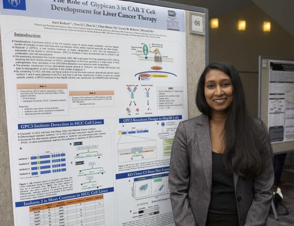
Soon after Aarti began her graduate studies at the Mayo Clinic in Rochester, Minnesota, she learned about an IRP researcher doing cutting-edge work on CAR-T cells, a type of re-engineered immune cell that scientists can tweak in order to treat a variety of cancers. With the blessing of her mentor at Mayo, she came to the NIH to work with senior investigator Mitchell Ho, Ph.D., on a new type of CAR-T cell therapy for liver cancer.
“It’s been a really cool experience because I’ve gotten to do exactly what I want to do,” Aarti says. “I get to use some of our new cell models from the Mayo Clinic here at NIH to test the therapies that we’re building.”
CAR-T cells are able to kill cancer cells by identifying a particular protein on the cancer cells, which triggers the CAR-T cells to begin their assault. Consequently, developing a CAR-T cell therapy requires identifying a molecule present on cancer cells that can signal CAR-T cells to attack. Aarti is specifically studying a molecule called glypican-3 that is active in more than 70 percent of liver cancer cells. So far, she has developed a set of tools, including cell models, that she believes she can use to learn more about glypican-3 and, ultimately, create and test CAR-T cells designed to attack cancer cells containing the protein.
“I think one of the best parts of being at the NIH is that it’s very diverse in terms of the science,” Aarti says. “Even though I’m a cancer person, I can go to a bacteria person and ask them a question. That kind of very broad research focus, but with the brightest minds, is an awesome part of being here.”
Hunter Schone: Peering Into Plasticity in the Human Brain
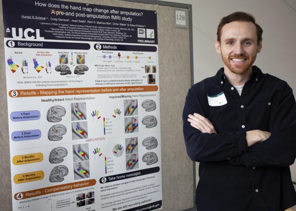
Hunter’s journey to the NIH had him cross the Atlantic Ocean not once but twice. Originally from Utah, he completed a Master’s degree at University College London (UCL) and is now pursuing a Ph.D. there while completing his dissertation research in the U.S. under the tutelage of IRP senior investigator Christopher Baker, Ph.D. Hunter and Dr. Baker share an interest in how experience changes the way our brains are organized, and one of the most extreme experiences a person can go through is amputation. A very small, very specific area in the brain is dedicated to controlling and processing input from each part of our body, and scientists like Hunter have long wondered how those areas change when the body part they have always been devoted to is no longer there.
To answer this question, Hunter recruited a patient who needed to have her right arm surgically removed due to a cancerous tumor and scanned her brain before and after the operation. He asked the patient to move each of her fingers while undergoing a functional MRI (fMRI) scan that recorded her brain activity, and during the post-amputation scans, he also asked her to attempt to move the “phantom” fingers she no longer possessed. Incredibly, even six months after the surgery, the parts of the patient’s brain dedicated to her missing fingers had changed very little. Hunter is now attempting to replicate this finding in additional patients.
“Our main conclusion is that we need to rethink the brain’s ability to actually change and completely re-organize these cortical maps,” Hunter explains.
“We see very limited changes in the brain’s representation of the hand despite losing an entire limb.”
In addition to providing basic insights into the brain’s ability to adapt to change and injury, Hunter hopes his work will aid the development of prosthetic devices that would enable patients to control mechanical body parts with their brains. This sort of cutting-edge work, he believes, can only be done at an extremely well-equipped institution like the NIH.
“The NIH has so many amazing resources,” he says. “When it comes to getting access to patients, getting access to scanning resources, having a collaborative network to work on these problems, I really don’t think I would have these things at any other institution.”
Bevin Blake: Examining the Effects of an Environmental Contaminant
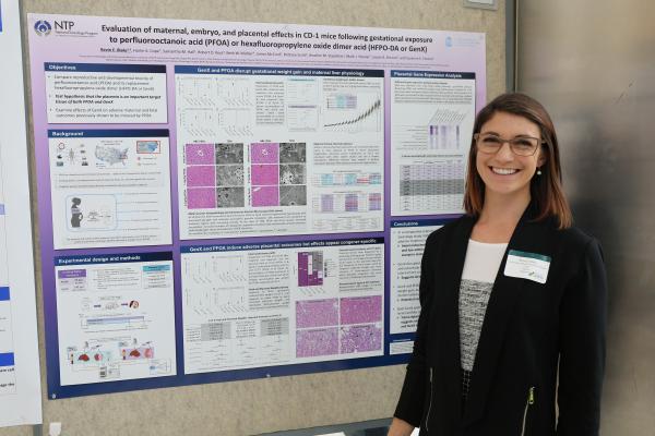
A graduate student at the University of North Carolina, Chapel Hill, Bevin is working with the IRP’s Suzanne Fenton, Ph.D., to learn about how certain chemicals released into the environment affect the health of mothers and their unborn children. Her dissertation project specifically focuses on a pair of chemicals, perfluorooctanoic acid (PFOA) and GenX, that are used to make products resistant to stains, grease, oil, and water. GenX was introduced to the market as a safer alternative to PFOA, which scientists had linked to adverse health outcomes in pregnant women.
Bevin began her project with the goal of determining exactly how PFOA affects the reproductive system and the developing fetus, but because GenX’s own safety profile has not been fully explored, she decided to examine its effects as well. The question of GenX’s effects on the body is all the more important because it has already contaminated North Carolina’s Cape Fear River, a major source of drinking water.
“There’s a pressing need to get some sort of baseline reproductive toxicity data on GenX, so since I was already going to be trying to understand how PFOA does what it does, I figured why not study GenX simultaneously to see if it does anything bad at all, and if it does, if it’s causing those effects in the same ways as PFOA.”
Bevin’s experiments showed that pregnant mice gained more weight after exposure to PFOA and GenX and that their livers also weighed more, a concerning finding because the liver is the main organ responsible for removing toxins from the body. What’s more, while GenX accumulated in the liver roughly 20 times less than PFOA, it harmed the liver just as much as an identical dose of PFOA. The continuing support of the IRP will help Bevin push this research further and learn even more about how these chemicals might affect human health.
“I have thoroughly enjoyed working at the NIH,” she says. “I feel that my research has been strengthened by access to some of the most cutting-edge tools and core facilities, so that has been really wonderful.”
John Fenimore: Investigating an Immune Assault on the Heart
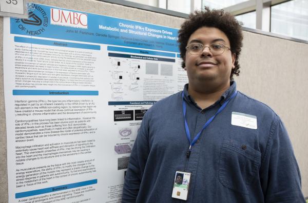
Unlike many of the students presenting their work at the symposium, John’s university and the IRP lab where he is performing his Ph.D. research are only about an hour’s drive apart. A graduate student at University of Maryland, Baltimore County, he is working with IRP senior investigator Howard Young, Ph.D., to study a mouse model that could yield insights into the relatively understudied population of men with lupus, an autoimmune disease in which a person’s immune system attacks his or her body.
“We’re digging down into relatively unmined soil,” John says.
Most lupus patients are female, but male patients are particularly prone to heart problems, and John is trying to understand why. Male lupus patients have elevated levels of an inflammatory molecule called IFN-gamma in their bodies, and the mice John studies have similar amounts of this molecule. In addition, he has discovered that the animals’ hearts show signs of damage and decreased function and that the mice have more immune cells called macrophages in their cardiac tissue than normal mice.
Now that John has an animal model that resembles the way lupus affects men, he can use it to test treatments that might help those patients. In fact, he has already seen positive results from two treatments that helped preserve heart function in the mice.
“Working in an NIH lab has been wonderful,” John says. “My mentor is very understanding about my need to balance my schoolwork with my time in the lab. I’ve found a lot of opportunities to explore my interests and take the reins of my research, I have access to a lot of resources, and I have a lot of freedom to use my time in a way that I think is most effective when it comes to my research.”
Rebekah Langston: Scrutinizing a Source of Parkinson’s Disease
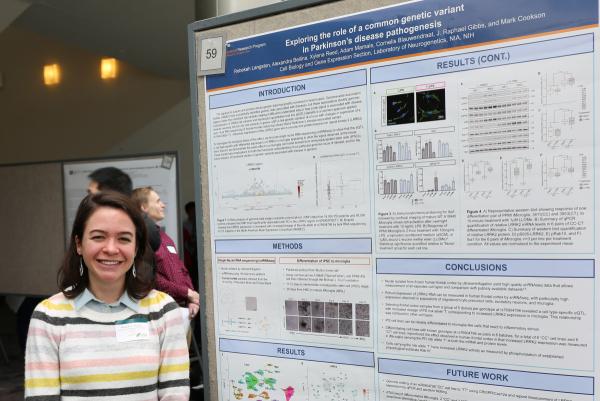
Rebekah is no stranger to the NIH, having worked here as a postbaccalaureate fellow after graduating college. Now in graduate school at the University of Arkansas for Medical Sciences in Little Rock, Rebekah opted to return to the NIH to work with IRP senior investigator Mark Cookson, Ph.D. She is specifically interested in a genetic variant in the LRRK2 gene that is associated with Parkinson’s disease in patients whose family members do not also have the illness, which destroys neurons related to movement.
By examining brain tissue donated by deceased individuals, Rebekah discovered that the LRRK2 gene is more active in the brains of people with that LRRK2 variant than in the brains of people without it, and this increased LRRK2 activity was confined to a specific type of brain cell called microglia, which remove damaged cells and defend the brain from bacteria and other pathogens. What’s more, the gene was similarly over-active in microglia grown in the lab that had the LRRK2gene variant compared to microglia without the variant. All together, Rebekah’s findings suggest that the genetic variant she is studying contributes to Parkinson’s disease through a mechanism that involves microglia, and she is now attempting to understand exactly what that process is.
“My project has been awesome and I’ve learned a lot,” she says. “It has really pushed me to learn a lot of different techniques. It’s also fun – I’m having a good time just because I get to do a lot of cool experiments.”
“Generally, the environment at NIH is unique because people aren’t worried about funding as much in the intramural program,” Rebekah continues. “I think that makes a big difference because we can explore avenues of research that other institutions might not be able to. We can do high-risk-high-reward experiments, which I think is really cool.”
Indeed, there may be no better place to begin one’s scientific career than in an IRP lab. The mentorship of seasoned, world-renowned scientists and the rich collection of resources available at the NIH will surely set these students up for long, successful careers full of important discoveries.
Subscribe to our weekly newsletter to stay up-to-date on the latest breakthroughs in the NIH Intramural Research Program.
Related Blog Posts
This page was last updated on Wednesday, July 5, 2023
