Colleagues: Recently Tenured
Meet your recently tenured colleagues: Grégoire Altan-Bonnet (NCI-CCR), Bevil Conway (NEI), Cari Kitahara (NCI-DCEG), Eros Lazzerini Denchi (NCI-CCR), Susan M. Lea (NCI-CCR), Sung-Yun Pai (NCI-CCR), Udo Rudloff (NCI-CCR), Leorey N. Saligan (NINR), and Lei Shi (NIDA).
GRÉGOIRE ALTAN-BONNET, PH.D., NCI-CCR
Senior Investigator and Head, ImmunoDynamics Group, Laboratory of Integrative Cancer Immunology, Center for Cancer Research, National Cancer Institute
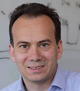
Education: École Normale Supérieure, Paris (B.S. and M.S. in physics); The Rockefeller University, New York (Ph.D. in physics)
Training: Postdoctoral fellow, Laboratory of Immunology, National Institute of Allergy and Infectious Diseases (2000–2005)
Before returning to NIH: Assistant then Associate member of the Programs in Computational Biology and Immunology, Memorial Sloan-Kettering Cancer Center (New York)
Came to NIH: In 2000–2005 for postdoctoral training; returned in 2015 as an Earl Stadtman Investigator in NCI
Outside interests: Hiking; sailing; cooking; having dinner parties with friends and colleagues (whenever we beat the pandemic)
Website: https://irp.nih.gov/pi/gregoire-altan-bonnet
Research Interests: Thanks to the pioneering work of Steve Rosenberg, colleagues in NCI’s intramural program, and others from all over the world, scientists have been able to harness the power of the immune system to attack tumors. The past 20 years have seen a flurry of novel approaches in cancer immunotherapy to boost leukocyte activation and tumor eradication. Yet we are lacking a systematic and quantitative understanding to propel the personalization, optimization, and generalization of cancer immunotherapies to all cancer patients.
To usher in such understanding, my group is developing experimentally validated quantitative models of the immune response—from signaling responses, to antigens and cytokines, to differentiation and proliferation of leukocytes, to killing of tumors. We engineered a novel robotic cell-culture system to map the combinatorial and dynamic complexity of leukocyte activation. We have also developed new quantitative methods (high-dimensional cytometry and spectral flow cytometry), machine learning, and computational modeling to identify hallmarks of successful immunotherapies (Science 370:1328–1334, 2020; DOI:10.1126/science.abb9847). [https://science.sciencemag.org/content/370/6522/1328]
Our current projects involve the multicellular coordination of immune responses against tumors and pathogenic infections. In collaboration with Nihal Altan-Bonnet’s group at NHLBI, we recently investigated how beta-coronaviruses—including SARS-CoV2, which causes COVID-19—use lysosomes to leave cells instead of using biosynthetic secretory pathways more commonly used by other viruses. Such highjacking of lysosomes by beta-coronaviruses explains some of the immune abnormalities observed in COVID-19 patients (Cell 183:1520–1535.e14, 2020). [https://pubmed.ncbi.nlm.nih.gov/33157038/] Our continued effort in systems immunology will help develop tailored immunotherapies against such pathogenic infections and tumors.
BEVIL CONWAY, PH.D., NEI
Senior Investigator, Sensation, Cognition, and Action Section, National Eye Institute

Education: McGill University, Montreal (B.Sc. in biology); Harvard Medical School, Boston (M.M.Sc.); Harvard University, Cambridge, Massachusetts (Ph.D. in neurobiology)
Training: Postdoctoral training at Harvard Medical School
Before coming to NIH: Associate professor in neuroscience, Wellesley College (Wellesley, Massachusetts) and principal research scientist at Massachusetts Institute of Technology (Cambridge, Massachusetts)
Came to NIH: In 2016
Outside interests: He is a visual artist.
Website: https://irp.nih.gov/pi/bevil-conway
Research interests: My group investigates the neural basis for visual perception and uses color as a model system for exploring how the brain processes sensory information. To better understand how the brain enables us to recognize faces, colors, objects, and places, my lab uses functional magnetic-resonance imaging (fMRI) and magnetoencephalography in human participants. These techniques use sensors around the head to noninvasively record blood-flow changes and tiny magnetic fields brought about by brain activity. The lab also does experiments in animal models to understand at a cellular and network level how the brain works.
Our work is organized around three broad approaches: 1) using fMRI in humans and nonhuman primates (NHPs) to investigate homologies of brain anatomy and function among these species and to test hypotheses about the fundamental organizational plan of the cerebral cortex in the primate; 2) using microelectrode recording in NHPs to show on a mechanistic level how populations of neurons drive behaviors such as perceptual decisions and categorization; and 3) doing comparative psychophysical studies in humans and NHPs as part of a program of neuroethology to understand the relative computational goals of perception and cognition in different primate species.
In addition to studies of vision, my team conducts experiments using auditory and combined audiovisual stimuli to understand common principles of sensory-cognitive information processing, and to determine how signals across the senses are integrated into a coherent experience. In recent work we discovered a substantial difference in how macaque monkey (Macaca mulatta) and human brains process auditory-pitch information (Nat Neurosci 22:1057–1060, 2019; DOI:10.1038/s41593-019-0410-7). [https://www.nature.com/articles/s41593-019-0410-7]
Other studies uncovered a universal pattern in how languages across the globe name colors (Proc Natl Acad Sci U S A 114:10785–10790, 2017; DOI:10.1073/pnas.1619666114). [https://www.pnas.org/content/early/2017/09/12/1619666114] In a third set of studies, we determined the color information carried by cells in the brain that are responsible for face recognition to test ideas about how the brain detects health and emotions of others. (eNeuro 2021; DOI:10.1523/ENEURO.0395-20.2020) [https://www.eneuro.org/content/eneuro/early/2021/01/13/ENEURO.0395-20.2020.full.pdf] and Nat Commun 10:3010, 2019; DOI:10.1038/s41467-019-10073-8). [https://www.nature.com/articles/s41467-019-10073-8]
And finally, our team recently used brain imaging to decode what colors people see. The study also revealed correlations between neurological signals and patterns in color naming. (Curr Biol 31:1–12, 2020; DOI:10.1016/j.cub.2020.10.062) [https://www.cell.com/current-biology/pdf/S0960-9822(20)31605-5.pdf]
CARI KITAHARA, PH.D., NCI-DCEG
Senior Investigator, Radiation Epidemiology Branch, Division of Cancer Epidemiology and Genetics, National Cancer Institute

Education: University of Michigan, Ann Arbor, Michigan (B.S. in biology and anthropology/zoology); Johns Hopkins Bloomberg School of Public Health, Baltimore (M.H.S. and Ph.D. in cancer epidemiology)
Training: Postdoctoral training at NCI-DCEG
Came to NIH: In 2007 for training; became a tenure track investigator in 2015
Outside interests: Playing with her two boys (ages 3 and 6); riding her Peloton bike; traveling (except during a pandemic)
Website: https://irp.nih.gov/pi/cari-kitahara
Research interests: My research focuses on 1) cancer risks in people—medical personnel as well as patients—who are exposed to diagnostic and therapeutic radiation and 2) the etiology of thyroid cancer, which is one of the most radiosensitive tumors.
Within the U.S. Radiologic Technologists Study, [https://dceg.cancer.gov/research/who-we-study/cohorts/us-radiologic-technologists] the largest cohort of radiologic technologists in the world, I demonstrated that medical staff who perform nuclear medicine procedures and fluoroscopically guided interventional procedures receive doses of radiation that are several times as high as those performing general radiologic procedures. Increased doses are associated with adverse health outcomes. These findings led to efforts to reinforce and improve personal protective equipment and occupational monitoring, and to informed regulatory decisions for occupational dose limits in the United States.
I also investigate risks to patients from nuclear medicine procedures. I led an international effort to extend the follow-up of the largest international cohort of patients with hyperthyroidism. My analysis of those data resulted in a report establishing that radioactive iodine treatment increases cancer risks in these patients (JAMA Intern Med 179:1034–1042, 2019; DOI:10.1001/jamainternmed.2019.0981). [https://jamanetwork.com/journals/jamainternalmedicine/fullarticle/2737319] Because of the potential for influencing treatment recommendations and patient preferences, my findings have been widely discussed by medical professional societies and clinical guidelines committees.
My descriptive research has shown rising trends in the incidence of advanced thyroid cancer and thyroid cancer–specific mortality, providing support for a true increase in the disease in the United States (JAMA 317:1338–1348, 2017; DOI:10.1001/jama.2017.2719). [https://jamanetwork.com/journals/jama/fullarticle/2613728] My etiologic research, which focuses on large cohort studies and international consortia, has helped to establish obesity as a modifiable risk factor for thyroid cancer and as an important contributor to the rising incidence rates.
EROS LAZZERINI DENCHI, PH.D., NCI-CCR
Senior Investigator and Head, Telomere Biology Unit, Laboratory of Genome Integrity, Center for Cancer Research, National Cancer Institute
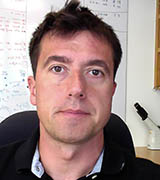
Education: University of Milan, Milan, Italy (B.S. and M.S. in biology); European Institute of Oncology, Milan (Ph.D. in molecular oncology)
Training: Postdoctoral fellow at Rockefeller University (New York)
Before coming to NIH: Associate professor, Department of Molecular Medicine, The Scripps Research Institute (La Jolla, California)
Came to NIH: In 2018 as a Stadtman Investigator; became tenured as a senior investigator in March 2021
Outside interests: Sailing and cycling
Website: https://irp.nih.gov/pi/eros-lazzerini-denchi
Research interests: Chromosome endcaps, called telomeres, shorten as one ages. I am interested in the mechanisms by which telomeres protect chromosome ends and their deregulation in aging and in pathologies such as cancer. My lab studies telomere-associated proteins to define their role in preventing the activation of the DNA damage-response pathway and suppression of end-to-end chromosomal fusions.
A complementary study in our lab is focused on the in vivo consequences of telomere dysfunction. Telomere length homeostasis plays a critical role in cellular and organism survival. However, in humans, this process is inefficient, and progressive telomere shortening is observed in tissues with a high cellular turnover. In the lab, we probe the role of telomere shortening in aging and tumor onset using mouse models that recapitulate telomere dysfunction in stem cells and other cellular compartments.
Our most recent work revealed that embryonic stem cells could survive and proliferate in the absence of proteins that are usually essential for telomere protection. In this work, we found that embryonic stem cells possess an alternative mechanism of telomere protection triggered by the induction of genes typically used only during the earliest stage of development to stave off unwanted DNA repair. These findings reported on November 25, 2020, in Nature (Nature 589:110–115, 2021; https://doi.org/10.1038/s41586-020-2959-4) [https://www.nature.com/articles/s41586-020-2959-4] might help explain the survival strategy used by some cancer cells to circumvent growth limits imposed by the natural shortening of telomeres that occurs as we age.
SUSAN M. LEA, D.PHIL., F.MED.SCI., NCI-CCR
Senior Investigator and Chief, Center for Structural Biology, Center for Cancer Research, National Cancer Institute

Education: University of Oxford, Oxford, England (B.A in physiological sciences; D.Phil. in molecular biophysics); elected Fellow of the United Kingdom Academy of Medical Sciences (F.Med.Sci.)
Training: Postdoctoral training, Laboratory of Molecular Biophysics, University of Oxford, and at Linacre College (Oxford)
Before coming to NIH: Chair of Microbiology, Sir William Dunn School of Pathology, University of Oxford; Scientific Director, Central Oxford Structural Microscopy and Imaging Centre (Oxford)
Came to NIH: December 2020
Outside interests: Listening to Baroque music; hiking; cooking
Website: https://irp.nih.gov/pi/susan-lea
Research interests: As a structural biologist, I have pioneered the use of mixed structural methods to study host-pathogen interactions and other medically important molecular pathways. My laboratory uses and develops cutting-edge structural methods including cryoelectron microscopy and X-ray crystallography to define molecular mechanisms involved in health and disease.
My lab is focusing on different biological systems ranging from bacterial pathogenesis to human cell division. We are looking at how large, multiprotein complexes, often membrane-crossing, are assembled. In our bacterial pathogenesis studies, we often examine the large membrane-spanning complexes involved in bacterial movement and the secretion of bacterial proteins and toxins. In our research on human systems, we examine how serum-resident protein cascades act in immune responses and coagulation; how centrosomes assemble during mitosis; and how a variety of membrane proteins function in protein and membrane maturation, as transporters or cellular receptors. We use multiple biophysical methods to address the questions we pose about these challenging systems, and we often develop software and experimental methods to allow us to obtain the answers we seek.
We have spent many years trying to understand the atomic details of the bacterial flagellum, the long appendage that many bacteria need to move through a fluid environment and cause disease. A flagellum is a multiprotein motor powered by the movement of ions across the bacterial membrane, an incredibly complex protein nanomachine. Our recent work has illuminated many previously unknown aspects of the machine and has revealed how the bacteria tightly intermesh the protein components with the membrane itself to contain the rapidly rotating motor. Building a clearer picture of these disease-causing machines will open opportunities for novel antibacterial agents in this age of antibiotic resistance. (References for research: 1) Accepted Nat Microbiol, bioRxiv 2020.12.05.413195, 2020; DOI:10.1101/2020.12.05.413195; https://www.biorxiv.org/content/10.1101/2020.12.05.413195v1; 2) Nat Microbiol 5:1553–1564, 2020; DOI:10.1038/s41564-020-0788-8; https://www.nature.com/articles/s41564-020-0788-8?proof=t; 3)
Nat Microbiol 5:966–975, 2020; DOI:10.1038/s41564-020-0703-3) https://www.nature.com/articles/s41564-020-0703-3
SUNG-YUN PAI, M.D., NCI-CCR
Senior Investigator and Chief, Immune Deficiency Cellular Therapy Program, Center for Cancer Research, National Cancer Institute

Education: Harvard College, Cambridge, Massachusetts (B.A. in East Asian languages and civilization); Harvard Medical School, Boston (M.D.)
Training: Residency in pediatrics, Boston Children’s Hospital (Boston); clinical fellowship in pediatric hematology/oncology, Boston Children’s Hospital and Dana-Farber Cancer Institute (Boston); research fellowship in rheumatology and allergy immunology, Brigham and Women’s Hospital (Boston)
Before coming to NIH: Associate professor in pediatrics, Harvard Medical School; attending physician at Boston Children’s Hospital and Dana-Farber Cancer Institute, and co-director of the Gene Therapy Program, Boston Children’s Hospital and Dana-Farber Cancer Institute
Came to NIH: In 2020
Outside interests: Spending time with family; cooking; playing flute; reading both fiction and nonfiction
Website: https://ccr.cancer.gov/idctp/sung-yun-pai
Research interests: I am developing and testing tailored cellular therapies, including allogeneic hematopoietic stem-cell transplantation (for example, bone-marrow transplant) and gene therapy, for children and adults who have genetic diseases of the blood and immune system. One of my goals is to understand how individual genes and the type of chemotherapy given before cellular therapy influences immune reconstitution.
Babies with severe combined immunodeficiency (SCID) are born without T lymphocytes and typically die in infancy without cellular treatment. I led a multi-institutional study with the Primary Immune Deficiency Treatment Consortium showing that giving SCID patients transplants before the onset of infection was critical to survival, that some types of SCID have better immune correction than others, and that using chemotherapy improves correction of the immune system (N Engl J Med 371:434–446, 2014; DOI:10.1056/NEJMoa1401177). [https://www.nejm.org/doi/full/10.1056/NEJMoa1401177] This study and other work underlie the open trial I am leading at more than 40 institutions that involve randomizing patients with SCID to either of two chemotherapy regimens.
Autologous gene therapy uses the patients’ own cells, representing the ultimate personalized treatment. We re-engineered a viral vector that was successful in treating the X-linked form of SCID but caused leukemia. We showed in a clinical trial that the new vector was just as effective and safer, with no leukemias to date after 10 years (N Engl J Med 371:1407–1417, 2014; DOI:10.1056/NEJMoa1404588). [https://www.nejm.org/doi/full/10.1056/NEJMoa1404588] We now have a multi-institutional trial of gene therapy for this disease using low-dose busulfan to further improve immune outcome. My team is also working on developing cellular therapies for Wiskott-Aldrich syndrome and deficiency of dedicator of cytokinesis protein 8.
UDO RUDLOFF, M.D., PH.D., NCI-CCR
Senior Investigator, Rare Tumor Initiative, Pediatric Oncology Branch, Center for Cancer Research, National Cancer Institute
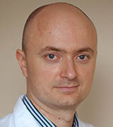
Education: Ruprecht Karls University School of Medicine of Heidelberg, Heidelberg, Germany (M.D., Ph.D.)
Training: Residency in obstetrics and gynecology at several different teaching hospitals in England; residency in general surgery, New York University School of Medicine and Albert Einstein College of Medicine (New York); fellowship in surgical oncology, Memorial Sloan-Kettering Cancer Center (New York)
Came to NIH: In 2009 as tenure-track investigator in NCI-CCR, first in the Surgery Branch (2009–2013), then in the Thoracic and GI Oncology Branch (2013–2018) and Rare Tumor Initiative, Pediatric Oncology Branch (2018–present)
Outside interests: Spending time with his nine-year-old daughter; reading; volunteering for the German-American Heritage Foundation; hiking
Website: https://irp.nih.gov/pi/udo-rudloff
Research interests: My laboratory concentrates on the discovery, translation, and early-phase clinical testing of novel therapies for patients with pancreatic or other solid-organ cancers. We have built a program that integrates phenotype-directed drug screening with target deconvolution (the process of identifying the molecular targets of active hits) and translational science to arrive at drug candidates that target novel essential mechanisms of cancer and yield candidates with favorable pharmacokinetic and toxicokinetic properties.
I led the efforts for the preclinical and clinical development of the first-in-class, small-molecule metarrestin, which effectively suppresses metastasis in pancreatic cancer and other solid-organ cancers. Metarrestin targets a marker of genome organization, the perinucleolar compartment, which is associated with the ability of cancer cells to metastasize. Orally administered metarrestin has favorable bioavailability and biodistribution across different species, achieves high intratumoral exposure concentrations, and has a predictable toxicity profile. We are currently enrolling patients in a first-in-human phase 1 study of metarrestin. (Sci Transl Med 10:441–455, 2018; DOI:10.1126/scitranslmed.aap8307). [https://www.ncbi.nlm.nih.gov/pmc/articles/PMC6176865/]
A second drug-discovery program focuses on tumor-associated macrophages, a major pro-tumor immune-cell population in many solid-organ cancers. Tumors typically co-opt macrophages to promote tumor growth and evasion from immune surveillance. In pancreatic cancer models, we found that the first-in-class synthetic host-defense peptide design RP-182 improved responses to chemotherapy and checkpoint blockades by binding to the innate immune checkpoint CD206 and targeting CD206-positive matrix-2 (M2)-like tumor-associated macrophages. (Sci Transl Med 12(530):eaax6337, 2019; DOI:10.1126/scitranslmed.aax6337). [https://stm.sciencemag.org/content/12/530/eaax6337]. RP-182 reprograms tumor-associated macrophages and increases cancer-cell phagocytosis and antitumor immune responses in various cancer models. Through several in silico (computer simulation), medicinal-chemistry, and structural-biology approaches we have recently replaced RP-182 with another small-molecule design that has improved pharmacokinetic features and is yielding a drug candidate for advanced preclinical studies.
We have been issued a patent for “Peptide-Based Methods for Treating Pancreatic Cancer” and a provisional patent for “Small Molecules with Selective Activity Against the M2 Phenotype of Macrophages.”
We have also found that pancreatic tumors with KRAS G12R mutations (present in 15–18% of pancreatic cancers) show increased response to the MEK inhibitor selumetinib in preclinical models. (Cancer Discov 10:104–123, 2020; DOI:10.1158/2159-8290.CD-19-1006). [https://pubmed.ncbi.nlm.nih.gov/31649109/] A phase 2 clinical trial, however, did not show tumor regressions in patients with advanced pancreatic cancer who were treated with a single-agent MEK inhibitor. We are currently evaluating, preclinically, the best possible MEK combination therapy for this patient subpopulation.
LEOREY N. SALIGAN, PH.D., R.N., C.R.N.P., F.A.A.N., NINR
Senior Investigator, Symptoms Biology Unit, and Acting Chief, Symptom Science Center, National Institute of Nursing Research
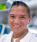
Education: Silliman University, Dumaguete City, Philippines (B.S in medical technology); Liceo de Cagayan University, Cagayan de Oro, Philippines (B.S. in nursing); Hampton University, Hampton, Virginia (M.S. and Ph.D. in family nursing)
Training: Postdoctoral fellow, Pain Research Unit, NINR
Before coming to NIH: Nurse practitioner, pulmonary and critical care specialist (Norfolk, Virginia); faculty, Family Nurse Practitioner Program, School of Nursing, Old Dominion University (Norfolk)
Came to NIH: In 2006 as nurse practitioner in NEI; did postdoctoral training in NINR’s Pain Research Unit (2007–2009); became associate clinical investigator in NINR in 2009; became tenure-track investigator in 2012
Outside interests: Running; traveling
Website: https://irp.nih.gov/pi/leorey-saligan
Research interests: I am exploring the nature and causes of fatigue in relation to cancer and its treatments. Fatigue is a common and debilitating condition that affects most cancer patients, impairing their health-related quality of life. To date, fatigue remains poorly characterized with no diagnostic test to objectively measure the severity of this condition. In addition, evidence has shown that the biology of cancer-related fatigue (CRF) is complex, and the condition may be caused by a cascade of biologic events in response to cancer and its treatments.
Acknowledging the individual variabilities in CRF, even among patients receiving similar treatments and having the same disease condition, my team observed that only a subpopulation of patients experience chronic fatigue after completing primary cancer treatment. The individual susceptibility to activate specific glutamate receptors seems to have a role in the chronic fatigue of a specific group of cancer patients months after completing their primary treatment. (Transl Psychiatry 8:article number 110, 2018; DOI:10.1038/s41398-018-0161-3). [https://www.nature.com/articles/s41398-018-0161-3] We used this discovery to conduct a proof-of-concept clinical trial [https://clinicaltrials.gov/ct2/show/NCT04141696] to investigate the antifatigue effects of inhibiting glutamate receptors using low-dose ketamine. This study is actively enrolling participants.
Recently, my group also found that cancer survivors carrying a specific polymorphism (Val66Met) of their brain-derived neurotrophic factor (BDNF) gene experienced less fatigue. Although Val66Met has been shown to greatly increase the risk for depression in healthy individuals, it did not affect depression symptoms in these cancer survivors (Transl Psychiatry 10:article number 302, 2020; DOI:10.1038/s41398-020-00990-4). [https://www.nature.com/articles/s41398-020-00990-4] This is an interesting discovery, considering that fatigue has always been associated with depressed mood. Using a mouse model, my team is exploring the unique brain networks associated with this specific BDNF polymorphism to help us understand how fatigue is perceived and experienced.
LEI SHI, PH.D., NIDA
Senior Investigator and Chief, Computational Chemistry and Molecular Biophysics Section, Molecular Targets and Medications Discovery Branch, National Institute on Drug Abuse
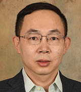
Education: Beijing University, Beijing (B.S. in biochemistry and molecular biology); Columbia University Medical Center, New York (M.A., M.Phil., and Ph.D. in pharmacology)
Training: Postdoctoral training in molecular recognition at Columbia University College of Physicians and Surgeons (New York) and in physiology, biophysics, and computational biomedicine at Weill Medical College of Cornell University (New York)
Before coming to NIH: Assistant professor, Department of Physiology and Biophysics, Weill Medical College of Cornell University
Came to NIH: In 2015
Outside interests: Playing and watching sports; enjoying wine; watching movies
Website: https://irp.nih.gov/pi/lei-shi
Research interests: Membrane proteins (MP) initiate intracellular signaling pathways, control the flow of energy and materials in and out of the cell, and thereby account for more than 30% of the human proteome and 40% of drug targets. My lab is investigating the structural basis of MP functions to advance the mechanistic understanding of key cellular processes. Using a combined computational and experimental approach, we identify and characterize the elements that determine the recognitions between MPs and their corresponding ligands, between MPs and their coupled proteins, and between MPs and the lipid environment. The integrated findings allow us to rationally develop small-molecule compounds for novel drug discovery.
Specifically, for G-protein-coupled receptors, our findings delineate the structural determinants of ligand selectivity and efficacy for dopamine receptors from both protein and small-compound perspectives. Our findings also describe the structural basis of allosteric mechanisms at these receptors. (Allosteric regulation is when a ligand, called an effector, binds to a protein site that is topographically distinct from a protein’s active site in which the activity characterizing the protein is carried out.) These mechanistic findings are being leveraged to identify novel lead compounds for both research and therapeutic purposes. For neurotransmitter transporters, we defined a novel allosteric mechanism of transport. Based on these findings, we and our collaborators discovered and optimized high-affinity allosteric inhibitors bound to serotonin transporters. Thus we have opened the door for clinical translation of allosteric inhibitors to reduce the side effects of current antidepressants.
This page was last updated on Monday, February 14, 2022
