Measuring Metabolism at the Speed of Light
Irene Georgakoudi’s WALS Lecture on Label-Free, High-Resolution Imaging
BY ANNELIESE NORRIS, NCI
Bioengineers build tools, always pushing the envelope on what is possible. They make technology better, faster, or easier to use. A new, advanced imaging technique can—quite literally—shine light on the potential to detect cancer earlier, assess disease progression, and inform better treatments.
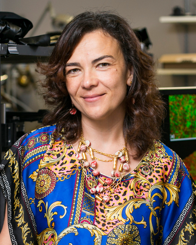
On May 24, 2023, Irene Georgakoudi, Professor of Biomedical Engineering at Tufts University (Boston, Massachusetts) delivered a Wednesday Afternoon Lecture Series (WALS) talk titled “Label-Free Monitoring of Metabolic Function with Micron-Scale Resolution: From Mitochondria to Humans” describing such advances.

CREDIT: TUFTS UNIVERSITY
Irene Georgakoudi is pushing the envelope with a new, advanced imaging technique that has the potential to detect cancer earlier, assess disease progression, and inform better treatments.
Celebrating the spirit of collaboration that runs deep in the bioengineering community, Georgakoudi’s visit to NIH was part of a two day mini-symposium that included poster presentations and lectures organized by the National Institute of Biomedical Imaging and Bioengineering (NIBIB).
“Her analysis techniques go way beyond the traditional approaches,” said NIBIB Director Bruce Tromberg in his welcome to Georgakoudi, who is also director of the Advanced Microscopic Imaging Center at Tufts. “In fact, she is the first in our field to generate unique mitochondrial information content from subwavelength structures; it’s a kind of functional super-resolution.”
Metabolic function as a measure of health and disease
Understanding and measuring how cells metabolize energy gives key information on the health of our busy mitochondria, which play an essential role in aging and the development of diseases such as cancer, aging, osteoarthritis, and neurodegenerative and cardiovascular diseases. However, current established methods to assess metabolic function such as radiographic, exogenous label, or mass-spectroscopy-based approaches can be invasive, often require contrast agents, or lack resolution.
“One of the challenges we’re particularly interested in addressing regarding [measuring] metabolic function is that metabolic changes are highly dynamic, some occurring in the context of milliseconds,” said Georgakoudi. “Others we would like to be able to monitor over weeks, days, and years.” Another challenge has been detecting different metabolic states often present in cells that lie right next to each other.
To address those challenges, the Georgakoudi lab pioneered the use of label-free, non-linear two-photon optical microscopy. This powerful imaging technique relies on lasers that emit very short pulses of long-wavelength, low-energy photons that penetrate into living tissue while minimizing tissue damage. When two of these photons are absorbed at the same time by NADH and FAD, they cause fluorescence. NADH and FAD are two naturally present co-enzymes associated with an array of metabolic pathways and mitochondrial status. The resulting 3D micron-scale resolution images are then analyzed for insights into the metabolic condition of the sample tissue.
Broad application for label-free, non-linear optical imaging
“We hope to change the paradigm of cervical precancer detection,” said Georgakoudi of a promising use for the optical imaging technology. Her team identified significant differences in mitochondrial organization and metabolic function between healthy cervical tissue and pre-cancerous lesions. She envisions a future where a two-photon microendoscope might be used in the clinic instead of biopsies to quickly and accurately diagnose precancerous lesions, which could then be treated on the spot.
Her new imaging approach has also revealed distinct mitochondrial organization in the living cells of patients with vitiligo, an autoimmune condition resulting in patches of skin-pigment loss. Further analysis has found that those lesions secrete inflammatory factors which may drive disease persistence. Because the optical imaging technique is non-destructive, Georgakoudi’s team is able to monitor how each patient responded to a skin grafting procedure over the course of several weeks.
Optical metabolic imaging may also give clues into life-extending pathways and lead to treatments for age-related diseases. Using the nematode Caenorhabditis elegans as a model, Georgakoudi and colleagues have demonstrated that worms deprived of a riboflavin transporter had better metabolic health and significantly longer lifespan, compared to controls.
Joint health can be assessed, too. In a mouse model for osteoarthritis, the optical imaging method detected very early metabolic remodeling changes in injured joint cartilage, before the onset of pain. The findings demonstrate how a better understanding of disease progression might improve how and when treatment is delivered.
As bioengineers do, Georgakoudi is poised to continue innovating, pushing the envelope of technology. “Our label-free two-photon optical metabolic imaging approaches reveal some very unique insights about metabolic function in living specimens and especially in humans” she said, but added, “We still have a way to go if we want to improve the way we diagnose and treat disease.”
To watch a videocast of Irene Georgakoudi’s lecture go to https://videocast.nih.gov/watch=46083 (NIH only).
Anneliese Norris, a postdoctoral fellow at the National Cancer Institute, is studying how signal interactions coordinate limb development. In her spare time, she enjoys reading and building with LEGO.
Bioengineers Collaborate, Innovate, Across NIH
BY MICHAEL TABASKO, OD
Wearable sensors, rapid diagnostics, biomaterials that speed up healing and deliver cancer vaccines, and more: On the day before Georgakoudi’s WALS lecture, a gathering of bioengineering experts assembled for what was billed as a “conclave” to “highlight novel biomedical imaging and engineering technology that they develop, and use, in their work,” according to Steven Zullo, a program officer with the National Institute of Biomedical Imaging and Bioengineering (NIBIB).
Zullo and fellow NIBIB Program Officer Afrouz Anderson organized the event, which also showcased poster presentations on the FAES Terrace.
Below are snapshots of the talks given by NIH intramural investigators. Watch the full VideoCast archive at https://videocast.nih.gov/watch=49750.
Accelerating healing
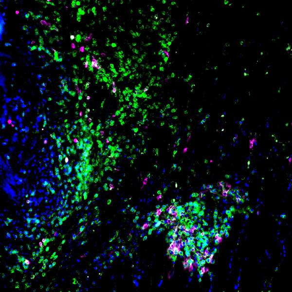
“I view engineering as an immunologist to develop new therapeutics,” said NIBIB Stadtman Investigator Kaitlyn Sadtler, Chief of the Section for Immunoengineering. She presented her work on how decellularized extracellular matrix (dECM)—a biomaterial that preserves a tissue's native environment, promotes cell proliferation, and provides cues for cell differentiation—produces a favorable cascade of immune system events and can improve how muscles heal after trauma.

CREDIT: KAITLYN SADTLER, NIBIB
Sadtler’s lab studies how the immune system responds to proregenerative biomaterials such as dECM after injury. Shown is injured mouse skeletal muscle (blue) treated with dECM. Molecules that modulate immune response and tissue healing, CD45 (green) and CD103 (pink), are shown responding to the injury site.
Controlling oxygen
Cells in a dish, or a drop, may experience vastly different concentrations of oxygen compared to their native environment. Those variations can affect scientific results. Nicole Morgan, chief of NIBIB’s Biomedical Engineering and Physical Science shared resource, is co-developing tools that control and measure oxygen in tissue cultures and help researchers conduct precision science.
Quantifying energy
Kong Chen directs the Human Energy and Body Weight Regulation Core at the National Institute of Diabetes and Digestive and Kidney Diseases. His team built a whole-room calorimeter at the NIH Clinical Center to measure human metabolic rate, which can help better understand diseases such as obesity. The Chen lab is using technologies such as metabolic chambers and wearable sensors for calculating energy metabolism, body composition, sleep, and physical activity in humans.
Innovating imaging
Children living with sickle-cell disease are at high risk for stroke, sometimes affecting cognitive regions of the brain. Manu Platt, director of NIBIB’s Biomedical Engineering Technology Acceleration Center, is using magnetic-resonance angiography and computer modeling to measure changes in blood-flow velocity. That information can indicate where stenosis and arterial damage is taking place. “We hope to establish a complete timeline of arteriopathy progression in the murine model of sickle-cell disease and confirm with histological analyses and test therapeutics,” said Platt.

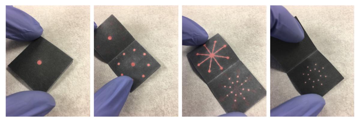
CREDIT: LICHEN XIANG, NINR
Shown is the paper origami multiplex sensor developed by Lichen Xiang which detects eight common gastrointestinal pathogens in stool for making rapid and accurate diagnoses in developing countries.
Creating lifesaving diagnostics
Diarrhea is a leading cause of childhood mortality in developing countries, but a low-cost diagnostic tool called the Paper Origami Multiplex Sensor has the potential to change that. Lichen Xiang, a research fellow at the National Institute of Nursing Research, developed the rapid, point-of-care test that detects eight common gastrointestinal pathogens in stool with over 98% accuracy and sensitivity.
Predicting neurodegeneration
The mighty mitochondria may play a role in brain health. Associate Scientist Qu Tian at the National Institute on Aging is measuring how impaired mitochondrial function in skeletal muscles can predict cognitive impairment. Her lab is studying how mitochondrial dysfunction is associated with biomarkers of Alzheimer’s disease and neurodegeneration. According to Tian, preliminary evidence has shown that exercise and dietary supplements may bolster mitochondrial health as we age.

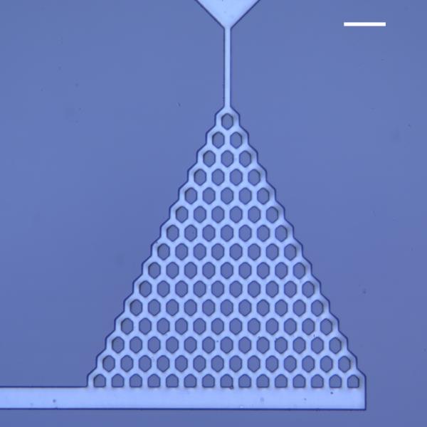
CREDIT: PANIZ REZVAN SANGSARI, NIBIB
Custom microfluidic device mimicking the branching network found in pulmonary microvasculature fabricated at BEPS’s Microfabrication and Microfluidics lab. Scale bar is 100 micrometers. Image taken with a microscope at BEPS.
Customizing devices
Biomedical engineer Paniz Rezvan Sangsari at NIBIB’s Microfabrication and Microfluidics Unit is using custom microfabricated devices to augment cell biology research. By patterning features down to 1 micrometer, bioengineers can model tissue environments to help researchers design realistic studies of cellular interactions. One such collaboration with the National Institute of Arthritis and Musculoskeletal and Skin Diseases helped researchers show how neutrophils navigate pulmonary arteries in patients with systemic lupus erythematosus.
This page was last updated on Tuesday, July 11, 2023
