neurons

Startling Snapshots of Science

Vibrant Images Push Forward Environmental Health Research
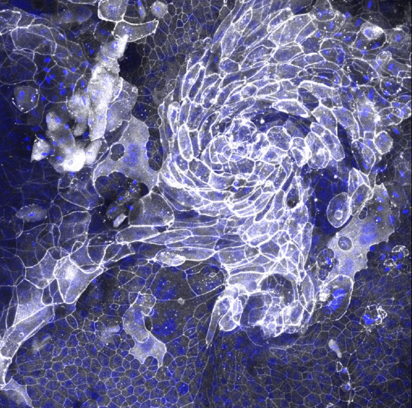
cell junctions in tracheal epithelial cells from a TLR5 knockout mouse

Scientists and artists obviously don’t spend their days doing the same thing, but that doesn’t mean the former can’t sometimes inadvertently be the latter. Take for example the 14 IRP research fellows who submitted images to the most recent edition of the Fellows Scientific Image Competition at NIH’s National Institute of Environmental Health Sciences (NIEHS). The competition, held every few years, shows that biomedical science can not only save lives, but also look good doing it.

“We are excited to showcase the work of our trainees using visual media, which reminds us there is art in science and showcases the important research we’re all engaged in at NIEHS,” says NIEHS Scientific Director Darryl Zeldin, M.D., who initiated the competition.

Tracking the Neuronal Response to Head Injury

New IRP Study Identifies Vulnerable and Resilient Brain Cells

man being treated for a head injury

Even with our thick skulls to protect our fragile brains, a hard enough blow to the head will still send our neurons into a tizzy. However, not all neurons are affected in the same way by a ‘traumatic brain injury,’ or TBI for short. By developing a way to see which neurons turn on a distress signal after a mild TBI, IRP researchers recently identified differences between neurons that die afterwards and those that are shaken up by the injury but manage to recover over time.

Antiviral Drug Stems the Spread of Parkinson’s-Promoting Protein

Study Shows Promise of New Treatment Approach in 3D Brain ‘Organoid’ Model

older man having difficulty eating soup

Our cells’ survival depends on their ability to take in the nutrients and other substances that they require. Unfortunately, this capacity is a double-edged sword, as cells can bring in both valuable resources and ticking time bombs. However, IRP researchers recently identified an existing drug that may be able to combat Parkinson’s disease by reducing cells’ penchant for snatching up the toxic proteins involved in Parkinson’s disease.

From Friend to Foe: When the Immune System Turns on the Brain

IRP Research is Exploring the Role of Immune Cells in Dementia

illustration of fire trucks rushing to put out a fire in the brain

If you visit a lab at NIH’s Center for Alzheimer’s and Related Dementias (CARD), you may find yourself surrounded by several robots hard at work nurturing the hundreds of sets of genetically modified stem cells that CARD scientists use to study the illnesses that give CARD its name. Many of these cells will be coaxed to mature into the neurons that power our movements, thoughts, and memories — but not all of them. Neurons have long received the lion share of dementia researchers’ attention, understandable seeing as the visible symptoms of Alzheimer’s disease are closely linked to a build-up of proteins — amyloid-beta and misfolded tau — that damage neurons. However, neurons aren’t the only brain cells involved in dementia.

Brain Pathway Amplifies Pain After Injury

Mouse Study Could Aid Development of Treatments for Chronic Pain

patient with shoulder injury talks with doctor

Getting hurt or sick is bad enough, but millions of patients around the world continue to experience pain or hypersensitivity even after their ailment resolves itself. Despite the prevalence of chronic pain, few effective treatments are available, especially ones without the potential for addiction that opioid medications carry. However, new IRP research has shown that suppressing the electrical firing of neurons in a certain brain area can alleviate injury-induced hypersensitivity in mice, providing a promising new target for treatments aimed at relieving chronic pain.

Gene Therapy Protects Neurons From Alzheimer’s Disease

New Approach Preserves Cognitive Abilities in Pre-Symptomatic Mice

diagram showing a virus entering a cell and delivering a gene into its nucleus

Cooks preparing traditional holiday feasts will surely find that a fire extinguisher can effectively quench an oven inferno, but it would probably be better if the oven never caught fire in the first place. Similarly, Alzheimer’s researchers have focused much more on putting out the biological fires scorching patients’ brains than on making brain cells ‘fireproof.’ A new IRP study in mice, however, suggests better results might be achieved with an approach specifically designed to make patients’ neurons more resilient.

Suppressing Neurons Curbs Withdrawal-Induced Pain Hypersensitivity

Insights From Mouse Study Could Aid Treatment for Opioid Use Disorder

man with neck pain

A headache or stubbed toe is annoying enough as it is, but for people in recovery from opioid use disorder, everyday aches and pains — and even sensations that would ordinarily not be painful at all — can be amped up to the point that they become quite distressing. A new IRP mouse study has helped scientists home in on the specific brain cells that might cause this phenomenon, providing a first step towards an intervention that could make it easier for people to stop using addictive opioid medications or illegal drugs like heroin.

Experimental Treatment Helps Neurons Recover From Damage

Mouse Study Could Lead to New Therapies for a Variety of Ailments

neurons firing

In most parts of your brain, the set of neurons you’re born with is what you’ve got for life — just like your fingers and toes, if you lose any, they’re not coming back. The body does have ways to encourage healing after a brain injury, but they are extremely constrained. However, by lending those natural systems a helping hand, IRP researchers have managed to dramatically boost regeneration and recovery of vision in mice with damage to the nerves that connect the eyes to the brain, an approach that could one day help people recover from other types of nervous system injuries as well.

Poster Session Showcases IRP Graduate Students

Event Includes In-Person Presentations for First Time Since 2020

people looking at posters at the 2023 NIH Graduate Student Research Symposium poster session

Three years after COVID-19 dramatically changed the way scientists and many others work, much of life in the NIH IRP has begun to resemble the way things were in February of 2020. This includes the return of in-person scientific poster sessions like the one that took place on February 16 as part of the 19th annual NIH Graduate Student Research Symposium. Nearly 130 graduate students conducting their Ph.D. research in IRP labs as part of NIH’s Graduate Partnership Program presented their progress at that poster session and its virtual counterpart held February 15.

The two poster sessions made it clear that IRP graduate students are essential contributors to the life-changing discoveries made at NIH, from using geckos to learn about human eye diseases to investigating how the immune system combats infectious invaders to exploring ways to improve cancer treatment. Keep reading to learn about some of the bright scientists-in-training who showed off their work during the two-day event.

IRP Scientist Shares Path From Stuttering to Science

Diversity-Focused NIH Program Helps Dr. Shahriar SheikhBahaei Investigate the Neuroscience of Voluntary Movement

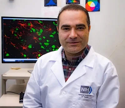
Dr. Shahriar SheikhBahaei

IRP neuroscientist Shahriar SheikhBahaei, Ph.D., first became aware of his stutter when he was 5 years old. Years later, his career would revolve around studying the biological roots of his speech impediment. But before he could start his own lab focused on how the brain controls voluntary movement, he needed a leg up from NIH’s Independent Research Scholars (IRS) program.