IRP Study Finds Brain Imaging Technique Could Aid Clinical Trials
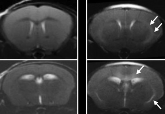
We all crave instant gratification, but unfortunately, most things worth doing take time. This certainly applies to evaluating treatments for many illnesses, as observable changes in patients’ symptoms can take weeks or months to appear. Fortunately, new NIH research has revealed that a particular way of non-invasively peering into the brain could help accelerate the development of treatments for a variety of rare neurological diseases.
The word ‘stroke,’ attributed to the idea of ‘a stroke of God’s hand,’ was first used in 1599 to describe the sudden seizure, paralysis, and brain damage that was previously called ‘apoplexy.’ It was a fitting analogy. Strokes, which affect nearly 800,000 Americans every year, hit suddenly and terrifyingly, with devastating consequences. Speed is critical to good treatment outcomes, but until recently very little could be done.
May is Stroke Awareness Month, a time to draw attention to the risks and symptoms of stroke and the new treatments that are helping people recover with fewer lasting effects. We recently spoke with IRP senior scientist Lawrence L. Latour, Ph.D., an expert on brain imaging who leads the Acute Cerebrovascular Diagnostics Unit, a unique partnership between the NIH Intramural Research Program and two hospitals in the metro, D.C., area: Suburban Hospital and Medstar Washington Hospital Center. The collaboration, launched in 2000, aimed to incorporate magnetic resonance imaging (MRI) in examinations of patients experiencing symptoms of stroke. This allowed the clinicians to diagnose patients more easily and then, through imaging at later time points, look at how patients responded to their treatments in order to learn ways to improve therapy.
Less Powerful Magnetic Fields Improve Heart and Lung Imagery
On November 8, 1895, a physics professor in Bavaria was working in his darkened laboratory when he noticed glimmers of light breaking through a piece of heavy black paper and lighting up a screen behind it. As he placed thicker and heavier items between the source of light and the screen, the light remained. That was the day Wilhelm Röntgen accidentally discovered x-rays and changed medicine forever.
As we celebrate World Radiology Day on the 128th anniversary of that discovery, medical imaging now allows people to see inside the human body with a clarity Dr. Röntgen scarcely could have imagined. Magnetic resonance imaging (MRI), in particular, has seen huge advances due to the development of bigger and stronger magnets. In contrast to that trend, IRP Stadtman Investigator Adrienne Campbell-Washburn, Ph.D., has instead combined better software and hardware with a less powerful magnetic field to create a new type of ‘low-field’ MRI that is particularly useful for taking pictures of the heart and lungs and for guiding minimally invasive procedures.
It’s been more than two years since the first outbreak of COVID-19 occurred in China. During that time, amazing scientific advances have dramatically altered prevention and treatment for the illness, including the development of remarkably safe and effective vaccines. However, even with widespread vaccination, scientists predict that the disease will continue to circulate in society indefinitely, with seasonal ebbs and flows like the flu.
As a result, even as COVID-19 vaccine shots rolled out by the hundreds of millions, numerous IRP researchers continued studying the disease and the virus responsible for it. Many of these projects have been funded by the NIH’s Intramural Targeted Anti-COVID-19 Program (ITAC), an initiative that provides IRP researchers with funding for research related to COVID-19. Over the past year and a half, ITAC has provided more than $12 million to support a wide variety of projects — more than can be covered in just one blog post. Read on to learn about just a handful of the many ways IRP researchers are contributing to the fight against COVID-19, and stay tuned next week for another blog describing even more ITAC-funded COVID research.
New Imaging Approach Improves Care for Men at Risk of Prostate Cancer
The National Academy of Medicine (NAM), first established in 1970 by the National Academy of Sciences as the Institute of Medicine (IOM), is comprised of more than 2,000 elected members from around the world who provide scientific and policy guidance on important matters relating to human health. Election to the NAM is considered one of the highest honors in the fields of health and medicine and recognizes individuals who have not only made critical scientific discoveries but have also demonstrated a laudable commitment to public service.
IRP senior investigator Peter Choyke, M.D., was elected to the NAM last year for his pioneering advances in imaging technologies for prostate cancer, which have improved diagnosis and treatment. Dr. Choyke, who directs the Molecular Imaging Branch at the National Cancer Institute (NCI), harnessed artificial intelligence to superimpose images from magnetic resonance imaging (MRI) scans onto images taken in real time using ultrasound, thereby enhancing doctors’ ability to non-invasively examine prostate tumors. This allows doctors to be more precise with their biopsies, thereby lowering discomfort and the risk of nerve damage when they take samples of the tumor. Better yet, if the images show no signs of cancer, patients can skip the invasive biopsy procedure entirely and just continue regular monitoring and checkups.
NIH Researcher Recognized for Contributions to Structural Biology Techniques
The National Academy of Sciences (NAS), established in 1863, is comprised of the United States’ most distinguished scientific scholars, including nearly 500 Nobel Prize winners. Members of the NAS are elected by their peers and entrusted with the responsibility of providing independent, objective advice on national matters related to science and technology in an effort to advance innovations in the United States.
IRP senior investigator Robert Tycko, Ph.D., was one of two NIH researchers elected to the NAS in 2020, an honor he hopes will give him the opportunity to help other scientists and improve the way science is done.
NIH Researcher Recognized for Advances in Brain Imaging
The National Academy of Engineering (NAE), established in 1964, comprises more than 2,000 peer-elected members who are among the world’s most accomplished engineers, including senior professionals in business, academia, and government.
IRP senior investigator Peter Basser, Ph.D., was elected to the NAE in February 2020. He directs the Section on Quantitative Imaging and Tissue Science and the Division of Translational Imaging and Genomic Integrity in the Eunice Kennedy Shriver National Institute of Child Health and Human Development (NICHD), where he studies how the structure and material properties of living tissues affect their function.
Future Physician-Scientists Spent a Year in IRP Labs
Many doctors not only treat patients directly, but also make valuable contributions to research that will improve medical care in the future. Each one of these talented ‘physician-scientists’ began his or her research career under the guidance of a more senior scientist. At the NIH, the Medical Research Scholars Program (MRSP) provides just such an experience to promising young medical students from all across the United States.
Study Results Could Help Improve Treatment for Alcohol-Related Problems
Your brain is always busy, even when you’re not thinking about anything. Scientists believe the way brain cells communicate with one another when the brain is in that ‘resting state’ might differ in individuals with certain diseases. In a recent study of this idea, IRP researchers found that resting state brain activity could effectively predict the severity of alcohol-related problems.
For Americans and others living outside the tropics, a mosquito bite is nothing more than an itchy inconvenience, but for billions of others, it can lead to a life-or-death battle with malaria. In some cases, the illness can wreak havoc on the brain. A new IRP study has used magnetic resonance imaging (MRI) technology to demonstrate that an investigational therapy can reverse that damage in mice.