A Weaker MRI Scanner Shows Its Strength
Less Powerful Magnetic Fields Improve Heart and Lung Imagery

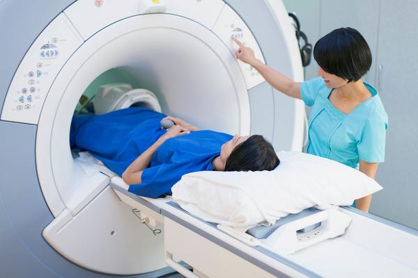
While most advances in magnetic resonance imaging (MRI) technology have relied on ever more powerful magnets, IRP researchers are showing that weaker magnets have surprising advantages.
On November 8, 1895, a physics professor in Bavaria was working in his darkened laboratory when he noticed glimmers of light breaking through a piece of heavy black paper and lighting up a screen behind it. As he placed thicker and heavier items between the source of light and the screen, the light remained. That was the day Wilhelm Röntgen accidentally discovered x-rays and changed medicine forever.
As we celebrate World Radiology Day on the 128th anniversary of that discovery, medical imaging now allows people to see inside the human body with a clarity Dr. Röntgen scarcely could have imagined. Magnetic resonance imaging (MRI), in particular, has seen huge advances due to the development of bigger and stronger magnets. In contrast to that trend, IRP Stadtman Investigator Adrienne Campbell-Washburn, Ph.D., has instead combined better software and hardware with a less powerful magnetic field to create a new type of ‘low-field’ MRI that is particularly useful for taking pictures of the heart and lungs and for guiding minimally invasive procedures.
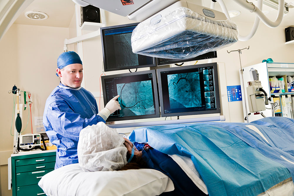
When Dr. Campbell-Washburn first came to NIH as a postdoctoral fellow in 2013, she joined a research team led by IRP senior investigator Robert Lederman, M.D., working to develop faster MRI technologies that could be used to guide heart catheterizations in real time. The procedure involves threading a thin tube through blood vessels and into the heart in order to make diagnostic measurements or treat damage. Unfortunately, MRI can heat up the catheters and metal tools used in the procedure to dangerous levels, so Dr. Campbell-Washburn's team wanted to make the system safer.
“If we really wanted these devices to be safe, I realized they needed to use a lower magnetic field strength because that was the only thing that would make them inherently safer,” she says.

Medical personnel use imaging technologies like x-rays and MRI to help them see inside the body during certain minimally invasive procedures. When those procedures use metal tools, the powerful magnetic fields generated by MRI scanners can be a problem.
At the same time, Dr. Campbell-Washburn and her colleagues were trying to improve MRI machines’ software with the goal of collecting MRI data more efficiently and converting it into higher quality pictures of our inner organs. After running several computer simulations, she and her colleagues determined that their tweaks to the system would not compromise an MRI’s ability to be used in heart catheterization, so in 2017, they decided to try implementing those changes in one of NIH’s MRI scanners.
This was a move that would have been impossible at any other institution, Dr. Campbell-Washburn says, due to the risk of modifying an expensive machine for the experiment. Fortunately, the risk turned out to be worthwhile. Not only did the lower-strength magnetic field make the machine easier to use for multiple MRI-guided medical treatments, but it made the machine itself more accessible as well.
“We’re really excited about this because the lower-field system is inherently less expensive than building a high-field system,” she explains. “They're easier to install, there are fewer requirements for how the room is configured, and they can be designed with wider openings for people with claustrophobia and obesity.”
“Our goal is not to replace high-magnetic field MRI; it’s to offer something different,” she adds. “You can imagine that if small hospitals had access to one of these systems, they could use them for many patients and just send patients who really need the high-field systems, such as for brain and spinal imaging, to the larger hospitals. Or, the scanners could be situated in different parts of the hospital closer to where patients are treated.”

Dr. Adrienne Campbell-Washburn
That vision is now moving closer to reality, as Dr. Campbell-Washburn has worked closely with a manufacturer that is now producing her low-field variety of MRI scanner. However, in spite of its benefits, radiologists were slow to accept Dr. Campbell-Washburn's low-magnetic-field MRI.
“There has been a drive over the past 30 years to adopt higher and higher magnetic fields to get better image resolution, and I was really going in the opposite direction,” she says.
However, acceptance came as Dr. Campbell-Washburn and her colleagues continued to improve the MRI machine and its software, including by integrating artificial intelligence and changing mathematical algorithms to improve clarity and improve the quality of their MRI system’s images. In fact, they discovered that their technology was actually better at scanning organs that are surrounded by or filled with air, such as the sinuses, lungs, and bowel.1
“We knew it would be better, but I don’t think we anticipated how much better,” Dr. Campbell-Washburn says.
The high-quality lung images her team has produced with its low-field MRI system have opened up new research avenues, including a study of COVID-19's long-term effects on the heart and lungs. The researchers are also developing more comprehensive lung imaging software to show the structure and function of expanding and contracting lungs.
"We certainly think that we've had a unique opportunity to study lung function with this MRI scanner that you can't get with any other technology,” Dr. Campbell-Washburn says. “We've been able to put a patient in the scanner and do an exam that involves comprehensive evaluation of both their heart and their lungs all in one place at the same time, which is just quite cool.”
This video shows scans of the lungs produced with the low-field MRI that Dr. Campbell-Washburn helped develop.
Now that she has proven the usefulness of her low-field MRI system in a few specific applications, Dr. Campbell-Washburn hopes to find additional uses for it in the years ahead.
“I work closely with interventional cardiologists, pulmonologists, and intensive care physicians to figure out new ways this technology can tell us something useful about patients,” she says. “A technology that is difficult to use or that doesn’t tell them anything new isn’t helpful. That’s really important to me.”
Subscribe to our weekly newsletter to stay up-to-date on the latest breakthroughs in the NIH Intramural Research Program.
References:
[1] Campbell-Washburn AE, Ramasawmy R, Restivo MC, Bhattacharya I, Basar B, Herzka DA, Hansen MS, Rogers T, Bandettini WP, McGuirt DR, Mancini C, Grodzki D, Schneider R, Majeed W, Bhat H, Xue H, Moss J, Malayeri AA, Jones EC, Koretsky AP, Kellman P, Chen MY, Lederman RJ, Balaban RS. Opportunities in Interventional and Diagnostic Imaging by Using High-Performance Low-Field-Strength MRI. Radiology. 2019;293(2):384-393. doi: 10.1148/radiol.2019190452.
Related Blog Posts
This page was last updated on Wednesday, November 8, 2023
