Colleagues: Recently Tenured
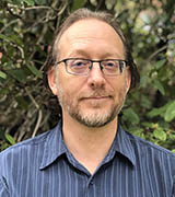
Meet your recently tenured colleagues: Andrea B. Apolo (NCI-CCR), Susan Harbison (NHLBI), Brandon K. Harvey (NIDA), Rosandra Kaplan (NCI-CCR), and Carlo Pierpaoli (NIBIB).
ANDREA B. APOLO, M.D., NCI-CCR
Senior Investigator and Chief, Bladder Cancer Section, Genitourinary Malignancies Branch, Center for Cancer Research, National Cancer Institute

Education: Lehman College, City University of New York (B.S. in chemistry and biochemistry); Albert Einstein College of Medicine, New York (M.D.)
Training: Residency in internal medicine at New York Presbyterian Hospital/Weill Cornell Medical Center (New York); fellowship in medical oncology, Memorial Sloan Kettering Cancer Center (New York)
Before coming to NIH: During her medical oncology fellowship, Apolo worked with Dean Bajorin at Memorial Sloan-Kettering and helped write and/or develop five therapeutic protocols for patients with bladder cancer.
Came to NIH: In 1996 as part of NIH’s Undergraduate Scholarship Program (1996–1998); returned in 2010 as assistant clinical investigator in NCI; in 2014 became a Lasker Clinical Research Scholar and chief of NCI’s Bladder Cancer Section, Genitourinary Malignancies Branch
Outside interests: Loves to spend time with her husband and their two boys, including hiking and biking together
Website: https://irp.nih.gov/pi/andrea-apolo
Research interests: I am interested in improving the treatment and survival of patients with genitourinary tumors. My team and I design and implement clinical trials to test novel agents for the treatment of urologic cancers. My primary research interest is in bladder cancer (urothelial carcinoma). In particular, we are developing new bladder-cancer therapies that use targeted agents including anti-angiogenesis compounds and inhibitors of Met receptors, which are essential for organogenesis and wound healing but are deregulated in some cancers. I test these targeted compounds individually or in combination with immunotherapies. We are also developing predictive and prognostic biomarkers in muscle-invasive and metastatic disease.
I led the bladder cancer study of avelumab that resulted in FDA approval of the drug for the treatment of advanced bladder cancer in people whose cancer has gotten worse when other therapies have failed (J Clin Oncol 35:2117–2124, 2017). Avelumab is an immune checkpoint inhibitor and blocks proteins called checkpoints that keep the immune system from working properly. The drug prevents programmed cell death protein 1 (PD1) on the surface of T cells from interacting with PD1 ligand 1 proteins on cancer cells. The T cells are then able to attack the cancer cells.
I also led a clinical trial testing the combination of a targeted therapy, cabozantinib, plus the checkpoint inhibitor nivolumab with or without ipilimumab, which led to the development of a phase 3 trial and FDA approval of this combination for patients with advanced kidney cancer.
SUSAN HARBISON, PH.D., NHLBI
Senior Investigator and Head, Laboratory of Systems Genetics, National Heart, Lung, and Blood Institute

Education: North Carolina State University, Raleigh, North Carolina (B.S. in aerospace engineering; Ph.D. in genetics)
Training: Postdoctoral fellowships in neuroscience, University of Pennsylvania (Philadelphia); postdoctoral fellowship in genetics, North Carolina State University
Other positions: Worked as an aerospace engineer for the Naval Aviation Depot (Marine Corps Air Station, Cherry Point, North Carolina) for eight years before completing her Ph.D. in genetics
Came to NIH: In 2012 as an Earl Stadtman Investigator in NHLBI
Outside interests: Hiking with her husband and their two Basenji dogs; powerlifting; knitting.
Website: https://irp.nih.gov/pi/susan-harbison
Research interests: People aren’t the only ones who sleep. Mammals, birds, fish, reptiles, amphibians, and invertebrates do, too. Poor sleep habits and sleep deprivation lead to ill effects on health and cognition. My lab and I are trying to better understand sleep and sleep disorders.
We are investigating the genetic networks underlying sleep and their interactions with the environment. We use Drosophila melanogaster (fruit fly) as a model organism, because fly sleep is similar to mammalian sleep. We have identified more than 1,500 genes associated with natural variations in sleep, many of which function in nerve-cell development. We also found that 250 genes account for variability in the circadian clock, including three genes that each contribute 1–1.5 hours to circadian cycle timing (J Biol Rhythms 36:239–253, 2021).
We are also studying how environmental changes—such as exposure to drugs, dietary changes, varying temperatures, and social isolation—can affect sleep in flies and humans. Daily sleep fluctuates in flies just as it does in humans. We identified genes that contribute to these intra-individual differences (Sleep 41:zsx205, 2018).
If sleep has a common purpose, then genes and gene networks affecting sleep are likely to be conserved across species. We bred flies for extreme sleep duration, producing flies that sleep for as long as 18 hours and for as little as 3 hours per day (PLoS Genet 13:e1007098, 2017). These populations enabled us to discover conserved genes for sleep duration and to develop community resources (Sci Rep 10:20652, 2020; G3 8:2865–2873, 2018). These genes may have similar functions in humans. For example, with colleagues at the University of Alabama at Birmingham, we found that a gene (dSdc) we uncovered in a Drosophila sleep study has a human homologue (SDC4) associated with sleep and resting metabolic rate in children (PLoS One 5:e11286,2010).
We hope that one day our research will lead to an understanding of the purpose of sleep, its role in human health, and the identification of targets and treatments for sleep disorders.
BRANDON K. HARVEY, PH.D., NIDA
Senior Investigator and Chief, Molecular Mechanisms of Cellular Stress and Inflammation Section, Integrative Neuroscience Branch, National Institute on Drug Abuse

Education: University of Rochester, Rochester, New York (B.S. in molecular genetics, M.S. and Ph.D. in neurobiology and anatomy)
Training: Postdoctoral training in NIDA
Came to NIH: In 2002 for training; became a staff scientist in 2006 and associate scientist in 2010; director of the Optogenetics and Transgenic Technology Core (2011–2016); in 2016, became a tenure-track investigator and chief of the Molecular Mechanisms of Cellular Stress and Inflammation Unit
Outside interests: Biking; kayaking; woodworking; gardening; and beekeeping
Website: https://irp.nih.gov/pi/brandon-harvey
Research interests: The Integrative Neuroscience Branch conducts research at the cellular, molecular, and systems levels to identify the neural substrates upon which substances of abuse act to produce long-term alterations in behavior and brain function. I lead the Molecular Mechanisms of Cellular Stress and Inflammation Section, in which we study the role of endoplasmic reticulum (ER) stress and inflammation in neuronal dysfunction caused by substance abuse or neurodegenerative diseases. The ER is a continuous membrane system within the cytoplasm of eukaryotic cells and performs such functions as protein synthesis and folding, lipid metabolism, and calcium storage. Dysregulation of ER is associated with neurodegenerative, muscular, and diabetic conditions.
We also identify and study the biology of secreted ER calcium modulated proteins (SERCaMPs) and Lys-Asp-Glu-Leu endoplasmic reticulum protein retention (KDEL) receptors; examine the influence of drugs of abuse and SERCaMPs on microglial activation; and develop genetic and pharmacological tools to monitor and modulate ER calcium.
In one study, we described a mechanism of cellular pathology linked to ER calcium depletion termed “exodosis,” which is observed in diabetes, stroke, Alzheimer disease, and cardiovascular diseases (Cell Rep 25:1829–1840.e6, 2018). In another study we identified a collection of small molecules that may play a therapeutic role in diseases associated with ER calcium dysfunction and exodosis (Cell Rep 35:109040, 2021).
We hope our work will lead to therapeutic strategies to restore ER function in a range of diseases.
ROSANDRA KAPLAN, M.D., NCI-CCR
Senior Investigator, Head of Tumor Microenvironment Section, Pediatric Oncology Branch, Center for Cancer Research, National Cancer Institute

Education: Connecticut College, New London, Connecticut (B.A. psychology and biochemistry); Dartmouth Medical School, Hanover, New Hampshire (M.D.)
Training: Residency in pediatrics at Boston Children’s Hospital (Harvard Medical School) and Boston Medical Center (Boston University School of Medicine, Boston)
Before coming to NIH: Assistant professor of pediatrics, Memorial Sloan Kettering Cancer Center and Weill Cornell Medical Center (New York)
Came to NIH: In 2010 as a tenure-track investigator
Outside interests: Loves staying active and spending time in nature with her family; loves to sing and before COVID enjoyed singing in an a cappella group
Website: https://irp.nih.gov/pi/rosandra-kaplan
Research interests: My lab and I are investigating how cancer spreads by avoiding immune detection and developing a structural matrix and its own blood supply that can support its metastasis to distant locations. Our work also aims to develop novel therapies that target a tumor’s ability to grow blood vessels and spread to other sites. We are detailing the earliest microenvironmental events in the metastatic cascade in hopes of translating our findings to the clinical setting.
When I was at Memorial Sloan Kettering Cancer Center and Weill-Cornell Medical College (New York), I worked with my research mentor to develop the concept of the pre-metastatic niche. We demonstrated that even localized tumors can prepare distant tissue sites for metastasis (Nature 438:820–827, 2005). We discovered that bone marrow–derived cells (such as immune cells known as myeloid cells) are recruited to future sites of metastasis and establish a receptive microenvironment for incoming circulating tumor cells. These specialized bone marrow–derived cells are present in circulating blood as well as in metastatic tissue of patients with pediatric and adult cancers. At NCI, my lab is focusing on the role of these cells in tumor progression, in neo-angiogenesis, in promoting immune evasion, and in regulating gene expression in the pre-metastatic niche.
In a recent study, we developed genetically engineered myeloid cells (GEMys) that deliver interleukin-12 (IL-12) to metastatic sites. We found that, in mouse models, chemotherapy with IL-12–GEMys reverses immune suppression and activates antitumor immunity (Cell 184:2033–2052.e21, 2021). GEMys might also be used to deliver other therapies to metastatic sites and be a treatment for late-stage malignancies and possibly other types of diseases that involve inflammation.
We are currently performing several studies to determine the trafficking of GEMys to tissue sites and their survival; the changes that occur with chemotherapy, radiation, and surgery; as well as differences in these cells in circulation and specific tissue sites. We hope our studies will lead to targeted therapies that can thwart the critical interaction of a tumor and its microenvironment as well as to myeloid-cell therapies that can potentially be used to modulate this microenvironment to limit metastatic progression.
CARLO PIERPAOLI, M.D., PH.D., NIBIB
Senior Investigator, Laboratory of Quantitative Medical Imaging, National Institute of Biomedical Imaging and Bioengineering

Education: University of Milan, Milan, Italy (M.D. and Ph.D. in neuroscience)
Training: Visiting fellow (1991–1995) and visiting associate (1995–1997), Neuroimaging Branch, National Institute of Neurological Disorders and Stroke (NINDS)
Came to NIH: In 1991 for training; became a visiting scientist and chief of NINDS’s Diffusion MRI Unit (1997–1999); became a staff scientist at the National Institute of Child Health and Human Development (1999–2016); in 2016 became a Stadtman Investigator in NIBIB
Outside interests: Sailing; hiking; cycling; cooking; stubbornly trying to give advice to his grown-up kids
Website: https://irp.nih.gov/pi/carlo-pierpaoli
Research interests: The goal of my research is to develop biomarkers that can characterize human anatomy and physiology across the lifespan and in disease using noninvasive imaging techniques, such as magnetic-resonance imaging (MRI). My lab and I aim to translate our research findings into effective clinical tools and to design a new role for radiology and imaging sciences in achieving noninvasive phenotyping with quantitative metrics that are ideally suited for inclusion in large, integrated databases.
We pioneered the clinical translation of diffusion tensor imaging (DTI), an MRI modality, that can noninvasively reveal information about tissue microstructures in the central nervous system. We performed the first DTI study of the human brain (Radiology 201:637–648, 1996). The MRI signatures we described in our early work are still used today by clinicians to interpret diffusion MRI changes in stroke.
In subsequent years, we worked consistently on making DTI metrics more biologically specific, reliable, and accurate. We proposed biomarkers that are informative of white-matter structure and organization, reversible brain ischemia, stroke, postnatal cortical maturation, and a type of nerve degeneration called Wallerian degeneration (Neuroimage 13:1174–1185, 2001). We worked on validating the accuracy of diffusion MRI to study brain connectivity (PNAS 111:16574–16579, 2014).
We have systematically addressed issues that make the use of potentially excellent biomarkers unreliable or even impossible in clinical applications. We investigated the effect of noise and artifacts such as subject motion, cardiac pulsation, or magnetic-field inhomogeneity on the computed metrics. We showed that these effects represent serious confounding factors in clinical applications, and we proposed a number of corrective strategies (Magn Reson Med 85:2696–2708, 2021).
We also developed TORTOISE, a software package used for processing diffusion MRI software data, and we have made it publicly available to the scientific and clinical communities for quantitative image analyses.
This page was last updated on Friday, January 28, 2022
