A Brain Signal Biomarker for Cerebral Palsy Treatments
Study Points to Potential Way to Improve Physical Therapy Interventions for Movement Disorder

Recent IRP research has linked communication between neurons in the brain to children’s performance on a movement task, suggesting a way to more quickly gauge the effectiveness of treatments for the movement disorder cerebral palsy.
It’s easy to take everyday activities like walking and typing for granted, but for people with cerebral palsy, any movement can be a struggle. Intensive physical therapy, starting at a young age, has long helped those individuals move more easily. Now, recent IRP research could provide a way to track how those and other treatments affect communication within the brain during movement, providing a tool that could help researchers more quickly evaluate which treatments work and which don’t.1
Movement disorders like cerebral palsy — CP for short — are a natural target of research focus for IRP senior investigator Diane Damiano, Ph.D., since she is one of the few — if not the only — physical therapists who leads a research team at NIH. A lot of her work focuses on designing and evaluating ways to help children with the condition, which she calls “the highest-burden chronic disease — beyond heart disease and diabetes — because it is quite prevalent, it affects the whole lifespan, and it can also present significant functional challenges.”
“Most children with CP can walk with or without assistance, while some require a wheelchair,” Dr. Damiano continues. “It’s quite a spectrum, and as you might imagine, their brains might function differently as well.”
Previous research exploring the brains of people with CP has mostly used magnetic resonance imaging (MRI), certainly a powerful tool but one with significant limitations. After all, placing people in a tight, noisy tube where any slight movement can compromise the images researchers get may not be the optimal approach for studying a movement disorder like CP. Electroencephalography (EEG), on the other hand, allows researchers to observe what is going on in the brain through electrodes placed directly on the head, which allows study participants to move much more freely.
“The ability to use EEG during functional movements is really improving exponentially in both the hardware and software, but it’s not for the faint of heart,” Dr. Damiano says. “It took us several years to feel confident in our data. The signals are very noisy and collection and processing of data requires a high level of technical expertise , but the incredible advantage is that you are to be able to do it while people are doing everyday tasks like walking or grasping objects rather than lying still or just moving a hand or a foot in the MRI.”

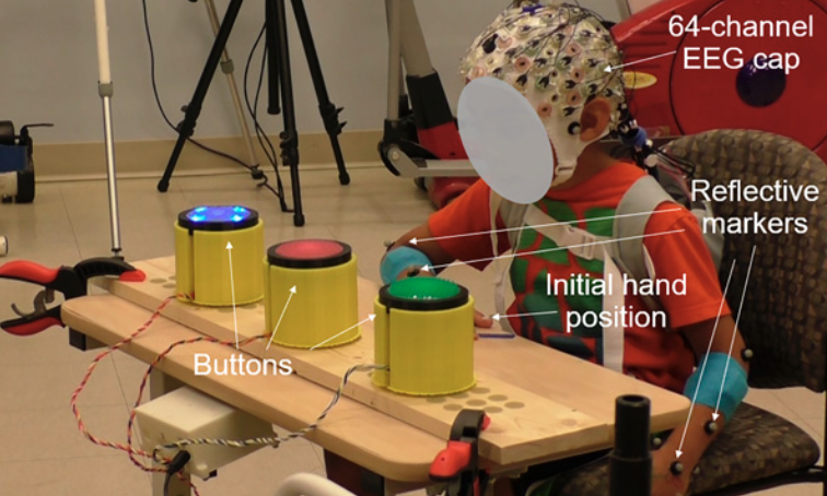
This photo shows a child participating in the study. The EEG records their brain activity in real time through 64 electrodes placed on each child’s head, while a computer can track and analyze the position of their arms and hands via ‘reflective markers’ placed on them.
In the newly published study, her research team monitored the brains of children with and without CP while they did a task requiring them to press one of three large buttons as soon as they could after the button lit up. Unsurprisingly, the children without CP did better at this. What’s more, as the researchers expected, there was an abnormally high amount of chatter between different parts of the brain when the children with CP moved compared to when the children without the condition moved, a measure called ‘connectivity.’ What the researchers had not predicted was that this connectivity was not elevated between the two halves of the brain, but rather within whichever half of the brain controlled the child’s non-dominant arm when he or she was using it. For instance, when right-handed children reached for the button with their left arm, the EEG showed more connectivity between different parts of the brain’s right half, which controls the left side of the body.
“It’s important that there’s connectivity in the brain, but what happens in CP is they have too much activation and too much connectivity,” Dr. Damiano explains. “When a child with CP goes to move, they activate too many muscles to do a task because their brain activation is too widespread compared to that in typically developing children. Brain effort equals muscle effort, so greater brain connectivity probably means they’re using too many muscles.”

Dr. Diane Damiano
The researchers were also particularly intrigued to see that when the children with CP were using their non-dominant arms, the ones who pressed the buttons more quickly after they lit up had less connectivity both within the half of the brain controlling the non-dominant arm and between the two halves of the brain. Tracking changes in that connectivity over time could provide a very useful way to assess the promise of new treatments for CP, particularly because it could potentially detect positive changes in the brain that emerge even before movement improves.
“The finding that more connectivity was associated with worse performance is important because now we can focus on reducing that connectivity,” Dr. Damiano says.
“Looking at a child’s movement alone cannot tell you how they’re using their brain,” she adds. “You have to look at their brain.”
Subscribe to our weekly newsletter to stay up-to-date on the latest breakthroughs in the NIH Intramural Research Program.
References:
[1] Lee SW, Bulea TC, Kline JE, Damiano DL. Dynamic Task-Related Changes in Electroencephalography Brain Connectivity During a Button-Press Task in Children with and Without Bilateral Cerebral Palsy. Brain Connect. 2025 May;15(4):162-174. doi: 10.1089/brain.2024.0096.
Related Blog Posts
This page was last updated on Thursday, July 10, 2025
