Taming Inflammation in the Intestines
IRP’s Warren Strober Breaks Down the Causes of Inflammatory Bowel Disease

IRP research has made important contributions to the understanding and treatment of inflammatory bowel disease.
For many, the holiday season brings expectations of delicious meals and treats with family and friends, but the nearly 1.6 million Americans who have inflammatory bowel disease (IBD) may need to skip these delights or endure serious digestive distress. It’s fitting, then, that the first week of December is Crohn’s and Colitis Awareness Week, an occasion that calls attention to the two conditions lumped together under the umbrella of IBD.
Of course, no awareness week is needed to remind IRP senior investigator Warren Strober, M.D., of the importance of learning more about those two conditions. An expert in how the immune system operates within the digestive system, Dr. Strober has spent decades looking for ways to provide relief for IBD sufferers.
“I decided in essence that it would be more important to study more common diseases of the digestive tract,” Dr. Strober says. “That’s why I turned my attention to inflammatory bowel disease.”

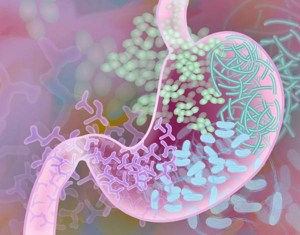
Dr. Strober’s research has revealed that inflammatory bowel disease often occurs because the immune system attacks the benign, native bacteria that live in our digestive system, known as the gut microbiome.
While Crohn’s disease and ulcerative colitis are both considered forms of IBD, they do have key differences. The inflammation caused by ulcerative colitis is limited to the intestines and colon, whereas Crohn’s disease can affect any part of the digestive tract, causing irritation and swelling of the tissue lining the walls of all organs involved, from the esophagus to the rectum. On the other hand, both conditions involve excessive immune responses to the native bacteria that normally exist harmlessly in our digestive tracts, and both are marked by distressing symptoms such as stomach pain and cramping, indigestion, constipation, and diarrhea.
Dr. Strober’s IRP laboratory focuses on the tissue that lines the walls of the digestive tract. That lining, called the mucosa, serves as a vital barrier that keeps harmful infectious organisms and environmental toxins out of our internal organs and blood. It is also the home of thousands of benign microorganisms, collectively called the microbiome, which play a vital role in maintaining a healthy immune system.
“We’re trying to find out how immune responses in the mucosal immune system can become abnormal and cause disease even though they’re responding to benign organisms that are normally found in a healthy gut,” Dr. Strober says. “The bacteria in the microbiome are not pathogens, so the disease occurs because the immune response is abnormal, not as a result of the bacteria it reacts to.”

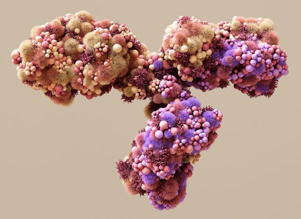
Dr. Strober’s research led to the development of an antibody that reduces the inflammation caused by Crohn’s disease and ulcerative colitis.
Dr. Strober’s laboratory began by studying immune system molecules called cytokines, which regulate inflammation. His was among the first laboratories to show that a particular type of cytokine, called interleukin-12 (IL-12), is important to the development of Crohn’s disease. Based on those findings, his laboratory created a mouse model designed to mimic the symptoms of Crohn’s disease and measured the level of IL-12 in the interior lining of the animals’ intestines, which turned out to be very high. Dr. Strober’s team also found they could stop IL-12 from doing its job in the body by using an antibody, a protein with a shape that allows it to stick to specific molecules. Not only did that antibody block IL-12’s effects, but it also lowered production of a second inflammation-inducing cytokine, IL-17, making its anti-inflammatory action even more potent.1 Ultimately, that antibody treatment was developed into a medicine called ustekinumab, now sold under the brand name Stelara® as a treatment for IBD.
After that important accomplishment, Dr. Strober and his team continued to dig deeper into the causes of IBD, seeking a genetic explanation for the excessive immune response behind it. They eventually decided to focus on a mutation in a gene that previous research had linked to Crohn’s disease, called NOD2. NOD2 normally causes immune cells to recognize bacteria in the gut and thus protects people from infection.
“Since the genetic NOD2 abnormalities occurring in Crohn’s cause loss of the NOD2 protein’s function, it seemed possible that these abnormalities cause Crohn’s disease by rendering individuals prone to infection,” Dr. Strober explains. “However, this is not likely because Crohn’s disease patients do not have more gastrointestinal infection than normal individuals and are, in fact, best treated with agents that lower resistance to infection.”
“To make a very long story short,” he continues, “we showed that when you have poor NOD2 function, other natural immune responses increase and attack our native bacteria, leading to the inflammation you see in Crohn’s disease. So NOD2 was a regulatory gene that kept the immune response in check.”
His group also studied another genetic mutation in a gene called ATG16L1, which controls the cellular clean-up process known as autophagy. Dr. Strober describes autophagy as “a sort of vacuum cleaner within the cell that scoops up debris and destroys it,” but it wasn’t clear why reduced autophagy increases risk for Crohn’s disease. The answer turned out to be that some of the molecular debris that piles up when ATG16L1 isn’t working activates other proteins in charge of creating inflammation.2
Having a faulty ATG16L1 gene isn’t necessarily a bad thing, though. Dr. Strober notes the mutation is present in about half the population and offers higher protection against infections. It’s only when the mutation is coupled with additional genetic risk factors that contribute to Crohn’s disease that it becomes problematic.
“We haven’t stopped there, of course,” Dr. Strober says. “There are over 200 different genes that can contribute to the risk not only for Crohn’s disease, but also for ulcerative colitis.”

A variety of genes influence the way the immune cells in the digestive tract respond to both harmful and benign bacteria.
The latest gene his group has been studying may be better known for its role in raising the risk of developing Parkinson’s disease; in fact, IRP senior investigator Andrew Singleton, Ph.D., recently won a prestigious award for establishing that link. In animal studies, Dr. Strober and his laboratory have shown that the same mutations in the LRRK2 gene that are associated with Parkinson’s disease are also linked with Crohn’s disease. Dr. Strober’s team discovered that when a faulty LRRK2 gene goes into overdrive, it can reduce autophagy and increase inflammation. This occurs, in part, due to the activation of an inflammatory molecule called NLRC4, which is normally only triggered by harmful pathogens like viruses and bacteria. However, hyperactive NLRC4 caused by an abnormal LRRK2 gene may attack the benign microbes that make up our microbiome, leading to intestinal inflammation.3

Dr. Warren Strober
Those studies spurred Dr. Strober’s team to form a collaboration with investigators at the Icahn Mount Sinai Medical Center in New York City, which treats many patients with Crohn’s disease. Together, the two groups are working on a treatment for Crohn’s that reduces the activity of the LRRK2 gene.
“If you have an inhibitor of LRRK2, you can block activation of NLRC4,” Dr. Strober explains. “We’re developing inhibitors that can be taken orally so they stay in the digestive tract and don’t inhibit LRRK2 in other parts of the body. Our collaborators send the inhibitors to us, and we test how well they work.”
That collaboration is ideal for Dr. Strober. While research hospitals larger than the NIH Clinical Center have access to more patients, the IRP provides a wealth of expertise and cutting-edge technology, he says.
“We have access to people who are steeped in knowledge about other kinds of technology, who can help you out,” Dr. Strober says. “No one person in today’s environment can know everything about doing research. We have to collaborate. This is one of the tremendous advantages of working at the NIH.”
Subscribe to our weekly newsletter to stay up-to-date on the latest breakthroughs in the NIH Intramural Research Program.
References:
[1] Mannon PJ, Fuss IJ, Mayer L, Elson CO, Sandborn WJ, Present D, Dolin B, Goodman N, Groden C, Hornung RL, Quezado M, Yang Z, Neurath MF, Salfeld J, Veldman GM, Schwertschlag U, Strober W; Anti-IL-12 Crohn's Disease Study Group. Anti-interleukin-12 antibody for active Crohn's disease. N Engl J Med. 2004 Nov 11;351(20):2069-79. doi: 10.1056/NEJMoa033402.
[2] Gao P, Liu H, Huang H, Zhang Q, Strober W, Zhang F. The inflammatory bowel disease-associated autophagy gene Atg16L1T300A acts as a dominant negative variant in mice. J Immunol. 2017 Mar 15;198(6):2457-2467.
[3] Takagawa T, Kitani A, Fuss I, Levine B, Brant SR, Peter I, Tajima M, Nakamura S, Strober W. An increase in LRRK2 suppresses autophagy and enhances Dectin-1-induced immunity in a mouse model of colitis. Sci Transl Med. 2018 Jun 6;10(444):eaan8162. doi: 10.1126/scitranslmed.aan8162.
Related Blog Posts
This page was last updated on Monday, December 4, 2023
