Colleagues: Recently Tenured
CHRISTOPHER BAKER, PH.D., NIMH
Senior Investigator, Section on Learning and Plasticity, Laboratory of Brain and Cognition
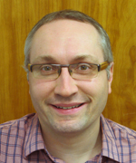
Education: University of Cambridge, Cambridge, England (M.A. in neuroscience); University of Saint Andrews, Saint Andrews, Scotland (Ph.D. in psychology)
Training: Postdoctoral training at the Center for the Neural Basis of Cognition, Carnegie Mellon University (Pittsburgh); postdoctoral associate, Department of Brain and Cognitive Sciences and McGovern Institute for Brain Research, Massachusetts Institute of Technology (Cambridge, Mass.)
Came to NIH: In 2006
Selected professional activities: Reviewing editor, The Journal of Neuroscience; academic editor, PLoS ONE
Outside interests: Trying to keep up with three very active children
Web site: https://irp.nih.gov/pi/chris-baker
Research interests: My research aims to better understand how the structure, function, and selectivity of the cerebral cortex change with experience or impairment, even in adulthood.
I am using brain-imaging techniques to explore three avenues of research. The first avenue concerns the nature of perceptual representations in the human brain, focusing on complex visual stimuli such as faces, bodies, scenes, and words.
In the second avenue of research, I am investigating how experience and learning change the neural and cognitive representations of sensory input. For example, my section is exploring the neural changes that underlie our enormous capacity to learn to recognize new objects and make fine-grained discriminations among them.
The third avenue concerns how the cerebral cortex adapts after damage to either the peripheral or the central nervous systems. For example, we are trying to determine the impact of macular degeneration or amputation on cortical function and how it relates to conditions such as phantom-limb pain.
Elucidating the nature and extent of cortical plasticity is critical for understanding how the brain functions throughout life.
AMY KLION, M.D., NIAID
Senior Investigator; Chief, Human Eosinophil Section, Laboratory of Parasitic Diseases
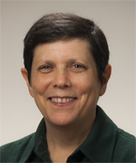
Education: Princeton University, Princeton, N.J. (B.A. biology); New York University School of Medicine, New York (M.D.)
Training: Residency in internal medicine at Johns Hopkins University (Baltimore); postdoctoral fellowship in NIAID’s Laboratory of Parasitic Diseases; postdoctoral fellowship in infectious diseases at the University of Iowa Hospitals and Clinics (Iowa City, Iowa)
Before coming to NIH: Assistant professor in the division of infectious diseases, University of Iowa College of Medicine (Iowa City)
Came to NIH: In 1989 through 1991 for training; returned in 1997 as a ßstaff clinician in NIAID’s Laboratory of Infectious Diseases; 2009 became a tenure-track clinical investigator
Selected professional activities: Member of the executive committee of the International Eosinophil Society; co-director of the Mali International Centers for Excellence in Research’s Filariasis Group
Outside interests: Spending time with family (including the dogs); traveling; and doing the New York Times crossword puzzle
Web site: https://irp.nih.gov/pi/amy-klion
Research interests: The Eosinophil Pathology Unit conducts basic and translational research related to the role of the eosinophil (a type of white blood cell) and eosinophil activation in disease pathogenesis. Our ultimate goal is to develop novel diagnostic tools and treatments for hypereosinophilic syndrome (HES) and other conditions associated with marked eosinophilia (a higher than normal concentration of eosinophils), including parasitic worm infections.
HES is a heterogeneous group of rare disorders in which eosinophils are responsible for the clinical manifestations. We are using genetic and immunologic tools to identify and characterize new subtypes of HES, such as an autosomal dominant form of familial eosinophilia that has been mapped to chromosome 5q31-33 and a rare cyclic form of eosinophilia associated with angioedema. Our clinical trials using targeted therapies, including imatinib mesylate and monoclonal antibodies to IL-5 and IL-5 receptors, have provided insight into the etiology and pathogenesis of HES variants.
Eosinophilia is common in human helminthic infections and may be associated with pathologic sequelae that mimic the clinical findings in HES, including tissue fibrosis and “allergic” manifestations. Furthermore, these manifestations may be produced or exacerbated by anthelminthic therapy.
We are assessing the safety and efficacy of chemotherapeutic agents targeting eosinophils (or their precursors) to prevent post-treatment reactions in loiasis, a filarial infection associated with dramatic eosinophilia after anthelminthic therapy. We are also exploring the role of the eosinophil in post-treatment reactions in other helminthic infections.
YOH-SUKE MUKOUYAMA, PH.D., NHLBI
(Legal name is Yosuke Mukoyama)
Senior Investigator; Laboratory of Stem Cell and Neuro-Vascular Biology
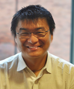
Education: Tokyo University of Science (A.B. in pharmacy studies); University of Tokyo (M.S. and Ph.D. in developmental biology)
Training: Postdoctoral training at the California Institute of Technology (Pasadena, Calif.)
Came to NIH: In 2006
Selected professional activities: Editorial board, Developmental Dynamics
Outside interests: Playing tennis and watching football games with family and friends
Web site: https://irp.nih.gov/pi/yoh-suke-mukouyama
Research interests: The goal of my research is to understand branching morphogenesis and patterning in organ development, especially in the vascular and nervous systems, which are often patterned similarly. My lab is using a combination of high-resolution whole-mount imaging, molecular manipulations, advanced genetic perturbations, and in vitro organ-culture techniques.
During angiogenesis, a primary capillary network undergoes intensive vascular remodeling and develops into a hierarchical vascular branching network. We have shown that sensory nerves determine the arterial branching pattern in embryonic skin. This alignment facilitates access to oxygen and nutrients for the nerves. At the molecular level, two distinct mechanisms underlie the sensory nerve-artery alignment: Nerve-derived vascular endothelial growth factor A controls arterial differentiation, and nerve-derived C-X-C motif chemokine 12 (also known as stromal cell-derived factor 1) controls vessel branching and alignment with nerves.
We have also shown that blood vessels influence nerve patterns in the embryonic heart. After the hierarchical vascular network is thoroughly covered with vascular smooth-muscle cells (VSMCs), sympathetic nerves extend along the network and innervate large vessels to control vascular tone and help regulate blood pressure. Coronary veins serve as intermediate conduits that guide distal sympathetic axon projections via the secretion of nerve growth factor (NGF) by coronary VSMCs. Subsequently, venous VSMCs downregulate NGF expression, and arterial VSMCs begin to secrete NGF, which stimulates the axons to innervate coronary arteries.
Our studies demonstrate a unique concept for understanding branching morphogenesis and patterning: Blood vessels and nerves take advantage of one another to follow the same path. Ultimately we hope to understand the developmental programs of branching morphogenesis and patterning in the vascular and neuronal branching networks and how we can restart these programs in diseased organs to generate healthy new networks. We are fortunate to be in a collaborative research environment that allows us to use core facilities and involve colleagues who have invaluable expertise.
VIPUL PERIWAL, PH.D., NIDDK
Senior Investigator; Chief, Laboratory of Biological Modeling, Computational Medicine Section

Education: California Institute of Technology, Pasadena, Calif. (B.S. in physics and mathematics); Princeton University, Princeton, N.J. (M.A. and Ph.D. in physics)
Training: Research physicist, Institute for Theoretical Physics, University of California, Santa Barbara, Calif. (1988¬1991); Member, Institute for Advanced Study, Princeton, N.J. (1991¬1993)
Before coming to NIH: Assistant professor, Physics Department, Princeton University
Came to NIH: In 2003
Outside interests: Reading fiction, biking, skiing, listening to classical music, and visiting art museums
Web site: http://1.usa.gov/1pu1Un4
Research interests: In my lab, we use mathematical models to understand and predict the functioning of human and animal metabolism and, more broadly, biological systems relevant to human disease. An important focus has been on insulin resistance, a major risk factor for several common diseases including diabetes, heart disease, high blood pressure, and some forms of cancer. The mechanisms underlying insulin resistance are not completely understood.
At the NIH, we developed the first mathematical model of mitochondrial metabolism, taking into account reactive oxygen species (ROS) and their control. Metabolism leads to the production of ROS; ROS signaling is important in normal cellular functioning. In oxidative stress, the body’s inability to detoxify and repair damage as fast as ROS are produced may be associated with insulin resistance. We developed a theoretical framework for modeling the dynamic development of the entire fat-cell size distribution. Ours was the first mathematical model of dynamic fat-tissue development. We continue to investigate how dynamic changes in fat tissue relate to insulin resistance and diabetes.
Additionally, we developed the first mathematical model of the formation and growth in the pancreas of the islets of Langerhans, the crucial endocrine micro-organs that maintain glucose concentrations. We also developed the first mathematical model of liver regeneration—in rats—and then showed that the model applies with almost no essential changes in humans who have been liver donors.
Using high-throughput biomedical datasets, we are gathering information on basic mechanistic molecular interactions and mechanisms. We are predicting the activity of transcription factors from DNA sequences, finding collective modes in protein structures from sequence alignments, and determining molecular interactions from single-cell data. Improving the understanding of disease processes should help in the development of effective treatments that do not have harmful side effects.
PETER WILLIAMSON, M.D., PH.D., NIAID
Senior Investigator; Chief, Translational Mycology Unit, Laboratory of Clinical Infectious Diseases
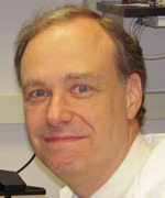
Education: Loyola University of Chicago (B.S. in chemistry, minor in mathematics); Boston University, Boston (M.D. and Ph.D. in medicine and biochemistry)
Training: Residency in internal medicine at Georgetown University Medical Center (Washington, D.C.); fellowship in infectious disease in NIAID
Before coming to NIH: Professor of medicine, pathology, microbiology, and immunology at the University of Illinois at Chicago
Came to NIH: From 1989 to 1994 for training; returned in 2009 to head NIAID’s Translational Mycology Unit
Selected professional activities: Editorial boards of Frontiers in Mycology and Journal of Mycology
Outside interests: Medical and medical educational activities in East Africa
Web site: https://irp.nih.gov/pi/peter-williamson
Research interests: The Translational Mycology Unit seeks to understand the role of host-pathogen genetics in the outcome of fungal infections. We use an array of methods including fungal genetics, cell biology, immunology, and population genetics to identify and validate weak points of the host-pathogen interface that might facilitate personalized therapeutic intervention.
My laboratory focuses on the AIDS-related pathogen Cryptococcus neoformans, which kills over a half a million people a year in regions of Africa and Asia, as well as Candida albicans, a major cause of bloodstream infections in the United States.
Specifically, we are interested in how a pathogen such as Cryptococcus undergoes genetic changes to convert itself from an environmental colonizer to a human pathogen. When the pathogen invades the host, changes in the expression of virulence factors optimize the acquisition of nutrients including copper, iron and glucose, paralyze the host immune response, and protect the fungus.
We use mouse modeling as well as high-dimensional data analyses of human isolates from people with and without AIDS to understand the fine coordination among genes that determine pathogenicity and suggest novel new treatment strategies. We have also used clinical genetic studies of patients with Candida albicans infections and mouse modeling to identify and study a human gene whose expression levels predict death during treatment.
In addition, we are recruiting an unusual cohort of patients to the NIH Clinical Center who do not have an underlying immunodeficiency but have acquired severe meningitis from the fungus for no apparent reason. In a trans-institute initiative with the National Institute of Neurological Disorders and Stroke, we are seeking to determine the nature of host defects that might have resulted in infection as well as to identify novel methods for immunomodulation of the disease to improve treatment outcomes.
This page was last updated on Tuesday, April 26, 2022
