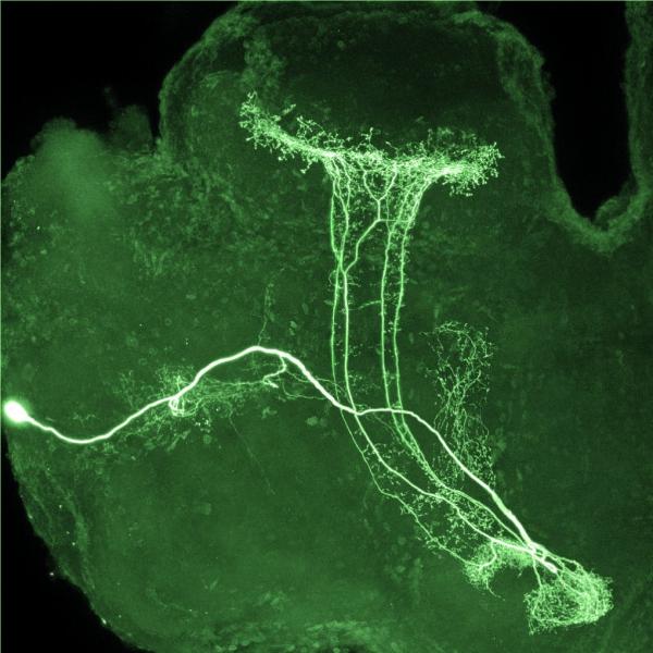
Sniffing Around Inside a Locust Brain

“Locusts provide a good simple model system for studying basic questions in neuroscience," writes Mark Stopfer, Ph.D., an Investigator at NICHD. “This image shows an olfactory interneuron innervating the beta-lobe of the locust brain. An intracellular electrode was used to record the electrophysiological responses of this neuron when odors were presented to the animal, and then to inject neurobiotin dye. The image was taken with a confocal microscope.”
Dr. Stopfer's group, the Section on Sensory Coding and Neural Ensembles, explores how brain mechanisms gather and organize sensory information to build internal representations of an animal's surroundings. By combining electrophysiological, anatomical, genetic, behavioral, computational, and other strategies, the group examines how fully intact neural circuits, driven by real sensory stimuli, process information. Learn more about his team's research.
Related Blog Posts
This page was last updated on Monday, January 29, 2024
