Postbac Life: A Week in an IRP Lab

I often use the microscope to check the quality of my immunohistochemistry stains and observe the state of fibrosis in lung tissue.
What does a postbac actually do in the NIH IRP? Maybe you have an image of someone mixing colorful chemicals together like a mad scientist (which sometimes isn’t too far from the truth).
Although I am not creating any diabolical concoctions, I am kept quite busy running tests to examine whether our treatments reverse the effects of lung fibrosis, a thickening and scarring of lung tissue. Here’s what a typical week looks like for me:
Monday
Measuring protein levels is important for showing what cellular activities are being restored in the lung. To study this, I weigh some mouse lungs that were flash-frozen in liquid nitrogen, specifically the left lung lobe (there are five lobes in a mouse lung). We need to know the lung’s weight to calculate the correct amount of chemicals to add later when we use the mass spectrometer to determine protein levels.
In the afternoon, I trim excess tissue off three of the other lobes. These lobes will be used to visualize the lung tissue’s structure by staining them and looking at them under a microscope. I place the lobes in plastic cassettes, which are then immersed in chemicals overnight.
Tuesday
I carefully remove the lung lobes from the cassette, place them in molten paraffin wax, and let the wax harden. The lungs are embedded in a block of wax so that we can more easily slice thin cross-sections of the lung (imagine trying to slice a <1 cm3 piece of tissue versus a large block of wax) using the microtome, which I mentioned in my previous post. I place the samples on glass microscope slides, which can be used later on (in this case, the next day) to stain and visualize various cellular components.
Wednesday

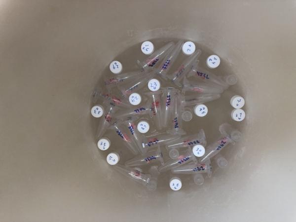
Mouse lung tissue sitting in liquid nitrogen, flash-frozen, ready to be processed to measure gene activity and protein levels.
Although the mass spectrometer shows us numerical values of protein levels, it is equally important to know exactly where in the lung the protein levels are higher or lower. We use immunohistochemistry (IHC) to visualize sites of fibrosis in the lungs. This technique relies on antibodies that flag proteins of interest — for us, ones involved in fibrosis. Using the slides prepared on Tuesday, I begin IHC staining by adding a primary antibody and incubating the slides in the fridge overnight.
We also study active gene expression levels by isolating RNA (which represents which genes are active) from the last lung lobe. I then convert it back into DNA by using a special enzyme that uses the RNA as a template to create a strand of DNA. We will use this DNA on Friday for gene expression analysis.
Thursday
I wash excess primary antibody off the slides and add the secondary antibody, which binds to the primary antibody. Our secondary antibody can then react with a chemical to stain the tissue brown in places where the primary antibody has latched onto the protein we’re looking for. We can view these stains under the microscope to see where protein levels are high or low and, consequently, if the fibrosis is reversed.
Friday
I use the DNA samples I obtained from Wednesday for quantitative PCR (qPCR) to quantify the levels of DNA in the lung tissue, which tells us how active a gene is. I usually run several plates of qPCR in a day. (It takes a lot of concentration and focus to pipet correctly into 96 wells per plate!) And just like that, the week is over.
Even though I have different tasks throughout the week, they are all closely related and reveal how the genes and proteins are acting on a molecular level. These aspects together help paint a more complete picture of how the drug we are testing works in an animal’s body. By performing these different aspects of gene expression analysis and tissue staining, we can see, either quantifiably or visually, whether the drug helps reverse fibrosis. We can then present these results to the scientific community so that, further down the line, our drug might be tested clinically and someday be used as a treatment in human patients with lung fibrosis. While I may play a small role, I am grateful for the opportunity to be a part of research that may one day help people suffering from this disease.
Related Blog Posts
This page was last updated on Monday, January 29, 2024
