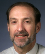
Colleagues: Recently Tenured
WILLIAM F. ANDERSON, M.D., M.P.H., NCI-DCEG
Senior Investigator, Epidemiology and Biostatistics Program, Biostatistics Branch

Education: University of Florida, Gainesville, Fla. (B.S. in chemistry); Tulane University School of Medicine, New Orleans (M.D.); Tulane University School of Public Health and Tropical Medicine (M.P.H. in epidemiology)
Training: Residency in internal medicine and fellowship in hematology and oncology at Tulane University School of Medicine; Cancer Prevention Fellowship Program at NCI
Before coming to NIH: Community-based hematologist and medical oncologist in West Monroe and Monroe, La.; founding board member of the Northeast Louisiana Cancer Institute (Monroe); staff physician and medical director of oncology unit at Glenwood Regional Medical Center (West Monroe); staff physician and medical director of oncology unit at Saint Francis Medical Center (Monroe); medical consultant for the Northeast Louisiana Tumor Registry (Monroe); clinical associate professor of medicine at Tulane University School of Medicine
Came to NIH: In January 1999 for training at NCI in cancer prevention and population-based science; in 2000 became a medical officer in NCI’s Division of Cancer Prevention; in 2005 became a principal investigator in NCI’s Division of Cancer Epidemiology and Genetics
Selected professional activities: Adjunct professor of pathology at the George Washington University School of Medicine (Washington, D.C.); adjunct professor of preventive medicine and biometrics at the Uniformed Services University of the Health Sciences (Bethesda, Md.)
Outside interests: Photographing birds and other wildlife
Research interests: After nearly 20 years as a private practitioner in hematology and medical oncology in rural northeast Louisiana, I developed an interest in cancer etiology, prevention, and descriptive epidemiology. In 1999, I came to NCI for training and stayed on to develop and lead a collaborative program in cancer-surveillance research. My experiences in private practice contributed to my broad interest in cancer—breast cancer in particular. I am trying to understand the effect of tumor heterogeneity on cancer etiology, pathology, gene expression, and survival, and how racial disparity and geographic variation affect cancer rates.
I supplement a hypothesis-driven approach to population-based science with biostatistical models such as mixture models, age-period-cohort models, and algorithms for replacing missing (or unknown) data and projecting future trends. My collaborators and I have made some intriguing discoveries that appear to characterize etiological heterogeneity.
We were the first to demonstrate that estrogen receptor (ER)–positive and –negative breast cancers have striking bimodal age distributions at diagnosis (that is, each of these cancers has two peak ages of onset instead of one).
We found that ER-positive tumors are bimodal with a late-onset peak near age 70 years, whereas ER-negative breast cancers are bimodal with an early-onset peak close to age 50 years. Although bimodal age-incidence patterns are acknowledged for malignancies such as Hodgkin lymphoma (a cancer of the lymphatic system), bimodality is not as well established for solid tumors such as breast cancer. Bimodality is important because it suggests cancer heterogeneity in an otherwise homogenous cancer population.
My success would not have been possible without the expertise, support, collaboration, and close interaction with colleagues in the Biostatistics Branch. The combination of advanced epidemiological and biological knowledge, biostatistical expertise, rich history of descriptive epidemiology, and tradition of stellar contributions on robust epidemiological methods places the branch in a unique position to be a world leader in future descriptive epidemiological studies and cancer surveillance research.
ROBERT NELSON, M.D., PH.D., NIDDK
Senior Investigator, Diabetes Epidemiology and Clinical Research Section, Phoenix Epidemiology and Clinical Research Branch

Education: Loma Linda University School of Medicine, Loma Linda, Calif. (B.S. in human biology, M.D.); Harvard University School of Public Health, Boston (M.P.H.); University of California, Los Angeles (Ph.D. in epidemiology)
Training: Residencies in internal medicine and in public health and general preventive medicine, Loma Linda University; epidemiology fellowship, Diabetes and Arthritis Epidemiology Section at NIDDK (Phoenix)
Before coming to NIH: Medical officer at the U.S. Naval Hospital (Okinawa, Japan); staff member at the Cleveland Clinic Foundation (Phoenix and Cleveland)
Came to NIH: From 1985 to 1988 for training; returned in 1994
Selected professional activities: Editorial boards of the American Journal of Kidney Diseases, Primary Care Diabetes, and Nephrology News and Issues
Outside interests: Hiking; biking; jogging; scuba diving
Research interests: Diabetes mellitus is the leading cause of kidney failure in the United States. My research focuses on kidney disease associated with type 2 diabetes. We study diabetic kidney disease in a Pima American Indian population near Phoenix that has a high frequency of type 2 diabetes. We are identifying risk factors for diabetic kidney disease and characterizing the functional and structural changes within the kidneys that occur with the development and progression of the disease. We are also seeking to identify new biomarkers of diabetic kidney disease that will help us detect it at an earlier stage and identify people who will respond best to various treatments.
We are conducting a clinical trial to determine whether treatment with a particular blood-pressure medicine will protect the kidneys from the damaging effects of diabetes. We are also examining gene expression in tissue obtained from kidney biopsies to identify molecular pathways responsible for kidney injury caused by diabetes.
The ultimate goal of our work is to characterize the clinical course of kidney disease in type 2 diabetes, identify the factors that increase a patient’s risk of developing this disease, and find better therapeutic approaches for its management and prevention.
MAHENDRA RAO, M.D., PH.D., NIAMS
Senior Investigator, Stem Cell Section, Laboratory of Stem Cell Biology; Director of NIH Center for Regenerative Medicine

Education: Bombay University, Mumbai, India (M.D.); California Institute of Technology, Pasadena, Calif. (Ph.D. in developmental neurobiology)
Training: Residency at Bombay University; postdoctoral training in neuroscience at Case Western University (Cleveland)
Before coming to NIH the first time: Associate professor at University of Utah School of Medicine (Salt Lake City)
First worked at NIH: May 2001 to October 2005 as senior investigator and chief of the Laboratory of Neuroscience in NIA
Before returning to NIH: Vice president, Regenerative Medicine, Life Technologies (Carlsbad, Calif.)
Returned to NIH: In August 2011 as director of the NIH Center for Regenerative Medicine
Selected professional activities: Working with international stem cell societies and regulatory agencies
Outside interests: Hiking; traveling
Research interests: My laboratory aims to use stem cells to improve our understanding of the biological processes controlling cell fate determination and tissue development. To accomplish this, we use stem cells to generate neurological disease models and to develop replacement therapies for neurodegenerative diseases. We hope versatile stem cells will become a source of replacement cells for damaged tissues.
One target for treatment with cellular therapies is Parkinson disease, which causes the death of nerve cells required for agile and controlled muscle movement. Symptoms of Parkinson disease include hand tremors and impaired walking. Alongside researchers at my previous position at Life Technologies, I developed methods to induce stem cell transformation into the type of nerve cells depleted in Parkinson disease. These nerve cells produce dopamine, a chemical signal that helps deliver the brain’s orders to the muscles. We derived such nerve cells from embryonic stem cells and adult induced pluripotent stem cells.
Other targets for treatment are peripheral neuropathies, particularly those caused by Schwann cell defects. We have developed methods to obtain a pure population of Schwann cells and are working with collaborators to develop a model to investigate myelination—the production of the electrically insulating material myelin, which forms a layer around the axon of a neuron. We will use this model to screen for small molecules that modulate the process of myelination and so may aid functional recovery.
PHILIP TOFILON, PH.D., NCI-CCR
Senior Investigator, Molecular Radiation Oncology Section, Radiation Oncology Branch

Education: University of Illinois, Urbana, Ill. (B.S. in physiology); University of Nebraska Medical Center, Omaha, Neb. (Ph.D. in pharmacology)
Training: Postdoctoral training in radiobiology in the Department of Neurological Surgery, University of California, San Francisco
Before coming to NIH: Professor of experimental radiation oncology and of neurosurgery at the University of Texas M.D. Anderson Cancer Center (Houston); faculty member at the Graduate School of Biomedical Science at the University of Texas Health Science Center (Houston); senior member of the Drug Discovery Department at the Moffitt Cancer Center (Tampa, Fla.); professor of oncologic sciences at the University of South Florida College of Medicine (Tampa, Fla.)
Came to NIH: From June 2001 until May 2006 as chief of NCI’s Molecular Radiation Therapeutics Branch; returned in June 2011 as an investigator in NCI’s Radiation Oncology Branch
Outside interests: Running; hiking
Research interests: In my lab, we are trying to understand the molecular determinants of cellular radiosensitivity—the susceptibility of cancer cells to the lethal effects of ionizing radiation. Our ultimate goal is to develop molecularly targeted agents that will make tumor cells more sensitive to radiation therapy. One project involves radiation-induced gene expression, which is thought to be the result of modifications in transcription [the copying of DNA into messenger RNA (mRNA)]. We recently found, however, that radiation induces changes in gene expression not by modulating transcription, but through regulating the translation of existing mRNAs into proteins. We are describing the mechanisms that mediate the radiation-induced translational control of gene expression and determining whether this process provides targets for tumor-specific radiosensitization.
We are also examining how the brain microenvironment affects the action of ionizing radiation on glioblastomas (GBMs), the most aggressive type of primary malignant brain tumors. This project involves irradiating human GBMs that have been transplanted into animal hosts: We implant human GBM stem-like cells—which have been isolated from surgical specimens and grown in vitro—into the brains of experimental mice. The goal of these studies is to use this model system to develop novel strategies for improving the treatment of GBMs.
YI-KUO YU, PH.D., NLM-NCBI
Senior Investigator, Quantitative Molecular Biological Physics Group, Computational Biology Branch

Education: National Taiwan University, Taipei, Taiwan (B.S. in physics); Columbia University, New York (M.A., M. Phil., and Ph.D. in physics)
Training: Postdoctoral training in the Department of Physics at Case Western Reserve University (Cleveland)
Before coming to NIH: Associate professor of physics at Florida Atlantic University (Boca Raton, Fla.)
Came to NIH: In May 2004
Outside interests: Playing classical guitar
Research interests: As a theoretical physicist working in biology, I have always been fascinated by the complexity in diverse organisms, all of which share a universal set of building blocks: water, ions, saccharides, fatty acids, amino acids, nucleotides, and other small molecules. However, our understanding of such complex systems is limited. My group is investigating on multiple levels—from the microscopic to macroscopic—how biomolecules function, interact with one another, and organize to form complex biological systems. Using theoretical physics and math, we are creating tools scientists can use to perform quantitative biological studies.
At the microscopic level, we have developed a method to accurately compute electrostatic forces and electrostatic energy between biomolecules. This method takes into account two major difficulties with biomolecules: their complex geometries and the presence of dielectrics (materials that do not conduct electricity well).
At the macroscopic level, we are exploring several areas in order to better understand how biomolecules organize to form complex systems. For example, we are looking at protein-protein interaction networks to clarify how proteins signal to each other and initiate DNA replication. My group has developed a mathematical framework that accounts for impaired communication between molecules, which, for example, can occur when proteases degrade proteins. We also use computational approaches to separate information from noise in massive biological data sets, thereby minimizing the misinterpretation of data.
In the realm of mass spectrometry (MS)–based proteomics, we have constructed computational tools for identifying peptides. MS can be used to determine a molecule’s chemical structure by measuring the mass-to-charge ratio of charged particles. Unfortunately, charged particles from outside sources can be detected and appear in the data set as well. Our computational tools, however, limit the appearance of these false data.
If you have been tenured in the past few months, The NIH Catalyst will be in touch with you soon to invite you to be included on these pages. We will ask for your CV and a recent photo, and then we will draft an article for your review.
This page was last updated on Monday, May 2, 2022
