Nobel Laureate Eric Betzig Shares “The Secret Lives of Cells”
Wednesday Afternoon Lecture Series Presentation in April 2019
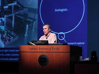
To create dynamic, precise images of cells, scientists have had to refine their microscopy methods to study them. Physicist Eric Betzig, a co-recipient of the 2014 Nobel Prize in Chemistry “for the development of super-resolved fluorescence microscopy,” delivered the lecture “The Secret Lives of Cells” on April 24, 2019. He described how he created sophisticated new microscopes that allow scientists to see living cells in action. Betzig has faculty appointments at the University of California at Berkeley (Berkeley, California) and at the Howard Hughes Medical Institute (HHMI) in Ashburn, Virginia.

CREDIT: CHIA-CHIA CHARLIE CHANG, NIH
Eric Betzig, who won the 2014 Nobel Prize in Chemistry “for the development of super-resolved fluorescence microscopy,” described “The Secret Lives of Cells” at an NIH WALS lecture recently.
The compound microscope was invented some 400 years ago, but until the 1980s, it was limited in its resolution and contrast. Throughout much of the 20th century, biologists created new techniques in genetics and biochemistry to study cellular dynamics: biochemical assays to break apart cells; molecular biology to determine the cellular genetic blueprint; and structural biology to understand the structure of cells. But these tools, while powerful, could not capture how various parts of living cells interacted with one another.
It wasn’t until the 1980s that the tables turned in favor of optical microscopy, thanks to the widespread availability of computers, lasers, sensitive detectors, and fluorescence-labeling techniques. These new technologies have given scientists the ability to understand the findings of genetics and biochemistry in the context of spatially complex and dynamic living systems at high spatiotemporal resolutions. Betzig described how he stepped into the field by chance.
In near-field optical microscopy, fluorescent molecules at the nanometer scale can illuminate unique optical properties. With proteins such as the green fluorescent protein (GFP), scientists can selectively label proteins and cells. Betzig’s thesis at Cornell University (Ithaca, New York), where he earned his Ph.D. in applied and engineering physics in 1988, was on near-field optics, an early form of super-resolution microscopy. After completing his doctorate, he went to work for AT&T Bell Laboratories (Murray Hill, New Jersey) on near-field optics. In 1989, a colleague at IBM demonstrated the ability to image single fluorescent molecules but only at temperatures near absolute zero. Using near-field optics, Betzig extended this to room temperature, and measured molecular positions to 20 times better than the resolution limit of standard microscopes. He won the National Academy of Sciences Award for Initiatives in Research for his work in 1993.
In 1994, he left Bell Labs and worked for other companies. In 2005 he and his old Bell Labs colleague Harald Hess created a prototype of the first super-resolution single-molecule localization microscope in Hess’s own living room. The invention would lead to seminal discoveries in biology. They then partnered with National Institute of Child Health and Human Development investigator Jennifer Lippincott-Schwartz, who had been working on using a photoactivitable form of GFP to visualize cellular-trafficking pathways, to first demonstrate their super-resolution photoactivated localization microscope, or PALM, in a small room in Building 32. In 2006, Betzig joined the Howard Hughes Medical Institute’s Janelia Farm Research Campus (Ashburn, Virginia) to further develop the technique. Lippincott-Schwartz joined HHMI in 2016.
Betzig has continued using PALM as well as other microscopes he and his group have developed since. His lattice light-sheet microscope, for example, produces high-speed 3D movies of cellular dynamics.
With a group at Stony Brook University (Stony Brook, New York), Betzig and his colleagues used this tool to observe cancer cellular migration in zebrafish ((Danio rerio), and with a group at MIT they created multicolor maps of the mouse visual cortex, quantifying the morphology of dendritic spines and the myelin sheaths that surround axons.
Most recently, Betzig and his HHMI colleagues have created a “Swiss Army Knife” mega-microscope to capture a wide range of spatiotemporal resolutions in a single instrument. They are building the first seven of them, at a cost of about $410,000 each; those and two more will be at Advanced Bioimaging Center at UC Berkeley that he is helping to establish. Through collaborations with Google and several biotech companies, the device would let data scientists make predictions and form conclusions using machine-learning and artificial-intelligence technologies. Each knife will generate about 20 terabytes of data a week.
In revealing the biological world at high precision, scientists can continue to uncover this secret life of cells.
“These technologies are finally able to allow us to understand the findings of molecular biology and biochemistry in the context of the dynamic cell,” Betzig said at the end of his talk. “I think that’s going to change the way we look at living systems in the future.”
To watch a videocast of Eric Betzig’s lecture (part of the Wednesday Afternoon Lecture Series) entitled “The Secret Lives of Cells” and presented on April 24, 2019, go to https://videocast.nih.gov/launch.asp?27460.
This page was last updated on Monday, April 4, 2022
