New Methods
Fluorescing Blobs Reveal Molecules
Seeing a lone molecule up close and personal just got faster and easier thanks to a new technique developed by scientists at NIDCD and NICHD.
“It’s very practical and very accessible,” said Bechara Kachar, head of the NIDCD Laboratory of Cell Structure and Dynamics and senior author on a study of the technique. “It doesn’t require further technical development, and the software is freely available. We hope that more researchers will take advantage of it.” (Proc Natl Acad Sci USA 108:21081–21086, 2011)
Even the most powerful light microscopes have limitations, and so to discern very tiny structures, scientists label them with “probes.” The fluorophore, a probe that absorbs and gives off light, works like a pedestrian wearing a reflective safety vest at night. If you see the vest, you can bet there’s a person underneath. If you see the fluorophore, you can safely assume a molecule is occupying the same spot.
Over time, as fluorophores emit light, they blink and bleach (lose color) like fireworks fading in the night sky. Kachar’s team wondered whether measuring these changes could help track individual molecules.

Uri Manor, NICHD
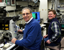
Bechara Kachar, M.D. (left), head of the NIDCD Laboratory of Cell Structure and Dynamics and senior author on the study about a new imaging technique, with first author Dylan T. Burnette, Ph.D., an NIGMS Pharmacology Research Associate (PRAT) Fellow working in the laboratory of Jennifer Lippincott-Schwartz (NICHD).
“Fluorescence images often look like blobs, since molecules overlap one another,” said the paper’s first author Dylan T. Burnette (NICHD).
So they made a technical and conceptual leap. First, they videorecorded a sequence of images in real time. Then, using copyright-free software (ImageJ: http://rsbweb.nih.gov/ij/) developed at NIH, they digitally subtracted from each image the subsequent image overlapping it. This process left the earlier image intact and let them see precisely where each molecule had been.
“If you take a photograph of the foliage of a tree in the summer, you cannot clearly visualize each leaf, although you know they are there,” said Kachar. “However, in autumn, when the leaves start falling individually, we can pinpoint the location of each leaf as it falls.”
Once they had detected each “fallen” or bleached molecule, they could calculate precisely its original location coordinates to make a comprehensive molecule map within a cellular structure.
“Bleaching-blinking-assisted localization microscopy, or BaLM, is a relatively simple and accessible new technique,” said Kachar. “Molecular information can now be attained where before there were only fluorescing blobs.”
This technique may help find the molecular hallmarks of diseases. “We always knew that all the data [were] there,” said Kachar. “We just had to reveal it.”
Imaging at NIH
The NIH intramural program has a long history of pioneering microscopic imaging techniques, such as the real-time picture processor, among the first computer hardware systems designed for imaging in the 1960s. Below are new techniques NIH researchers and colleagues at Howard Hughes Medical Institute’s (HHMI) Janelia Farm Research Campus (Ashburn, Va.) are developing. The tradition lives on, as demonstrated in a AAAS webinar featuring three NIH scientists on February 29, 2012, titled “Applying New Imaging Techniques to Your Research: Advice from the Experts.” (See http://bit.ly/zhpKUC.)
PALM: Photo-activated Localization Microscopy combines single fluorophores (single-molecule imaging) with controlled activation of these molecules for a composite image, providing “super” resolution at 10 to 20 nanometers, about ten times the size of an average protein; invented by Harald Hess and Eric Betzig, now at Janelia Farm, and further developed and exploited by Jennifer Lippincott Schwartz (NICHD) and others.
PALM applications: Two-color PALM, spt-PALM (single-particle tracking), iPALM (interferometric), and three-dimensional PALM have qualities beneficial for imaging certain cell types and depths; developed by NIH and HHMI.
iSPIM: The “i” is for “inverted,” a spin on SPIM, single-plane illumination microscopy, used for noninvasive imaging of living samples, such as Caenorhabditis elegans; see Y. Wu (NIBIB) et al. (Proc Natl Acad Sci USA 108:17708–17713, 2011)
MSIM: The “m” is for “multifocal,” a twist added to structured illumination microscopy that uses a multifocal pattern to do structured illumination microscopy to a depth of about 50 micrometers.
TED: Total-emission detection is a method that maximizes the probability of collecting all the scattered and ballistic light generated at the focal spot to optimize the signal-to-noise ratio about 10-fold; a spinoff is epiTED for live in vivo imaging.
Bessel Sheet Microscopy: This technology uses a thin sheet of light, akin to a barcode scanner, to peer inside single living cells, and combines high spatial resolution and temporal detail; efforts led by HHMI with samples from the NIH.
Special thanks to Catherine Galbraith (NICHD) and Hari Shroff (NIBIB) for helping to compile this list.
This page was last updated on Monday, May 2, 2022
