By IRP Staff Blogger
Monday, February 27, 2017

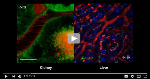
Roberto Weigert is a cell biologist who specializes in intravital microscopy (IVM), an extremely high-resolution imaging tool that traces its origins to the 19th century. What’s unique about IVM is its phenomenal resolution can be used in living animals, allowing researchers to watch biological processes unfold in organs under real physiological conditions and in real time.
By IRP Staff Blogger
Tuesday, March 1, 2016

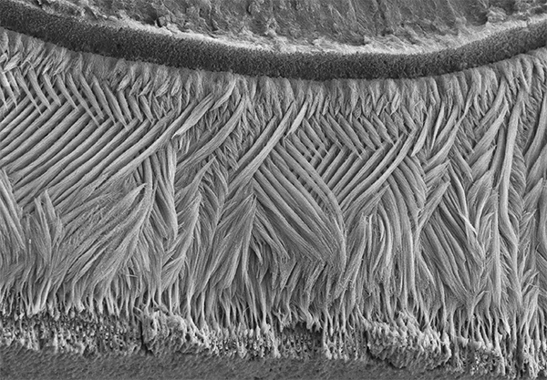
If you went out and asked folks what they’re seeing in this picture, most would probably guess an elegantly woven basket, or a soft, downy feather. But what this scanning electron micrograph actually shows isn’t at all soft: it is the hardest substance in the mammalian body—tooth enamel!
By IRP Staff Blogger
Tuesday, February 23, 2016
Scientists in the NIH Intramural Research Program (IRP) go to work each day in the biomedical equivalent of a candy store. The advanced equipment and shared tools readily available to researchers doing basic, translational, and clinical science in the IRP are unmatched anywhere else, which enables high efficiency and productivity within the IRP’s unique discovery model. Read more...
By IRP Staff Blogger
Wednesday, January 20, 2016
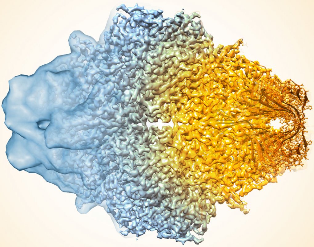
In the quest to find faster, better ways of mapping the structure of proteins and other key biological molecules, a growing number of researchers are turning to an innovative method that pushes the idea of a freeze frame to a whole new level: cryo-electron microscopy (cryo-EM).

By IRP Staff Blogger
Wednesday, December 2, 2015
Until recently, visualizing many of the processes underlying human diseases had been almost impossible. The NIH IRP’s role in developing technologies that can peer into human organs and cells is a key component of the Long-Term Intramural Research Program Planning Report and basis of sessions during this year’s #ResearchFest.
By Ashleigh LoVette
Monday, April 13, 2015
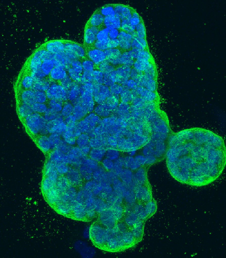
Monitoring cell movement. Examining the microenvironment of a tumor. Mapping a gene. Scientists at the NCI Center for Cancer Research (CCR)—the intramural research program at the National Cancer Institute—use a wide variety of microscopy techniques to observe and probe the otherwise invisible processes that drive cancer at the molecular level.

By IRP Staff Blogger
Wednesday, February 25, 2015
As part of a microscopy course at the National Center For Biological Sciences in Bangalore, India, Dr. Hari Shroff and Dr. Abhishek Kumar of the NIBIB IRP led a team of students in building a dual-view selective plane illumination microscope (diSPIM). It took them about eight hours to complete, but you can view the whole process in just four minutes in the timelapse video below:
By Sara Lioi
Sunday, November 16, 2014
As a postdoc in the Membrane Transport Biophysics Unit at NINDS, I’ve spent the past several years studying lysosomal pH. For me, that means spending a lot of my time in a pitch-black room, a room that I refer to as my cave.