IRP Research Challenges Long-Held Ideas About Muscle Structure
By Melissa Glim
Wednesday, January 19, 2022
It’s not every day an accidental observation overturns 100 years of biological knowledge. But that’s what happened when IRP Stadtman Investigator Brian Glancy, Ph.D., noticed something funny while reviewing high-definition 3D videos of muscle cells.
“To be honest, you could almost call this study an accident,” he says.
Dr. Glancy, who leads the National Heart, Lung, and Blood Institute (NHLBI)’s Muscle Energetics Lab, often uses the high-powered microscopes available through the NHLBI Electron Microscopy Core to study how energy is distributed through skeletal muscle cells — the ones that control voluntary movement — when they expand and contract.
Although he was focused on examining the cells’ energy-producing mitochondria, he could also see the other structures inside them, including the long, tube-like structures called myofibrils that are involved in muscle contraction. As he advanced the video and traveled down the length of the muscle, it looked to him like the myofibrils were changing shape.
By Michele Lyons
Thursday, March 14, 2019
Surrounded as we are with incredible technologies like supercomputers, MRI scanners, and smartphones, it's easy to forget that technologies viewed as antiquated today were once considered cutting-edge. Perhaps learning about some of the gadgets and technological concerns from NIH's past will help spark a greater appreciation for the wonderful gizmos that are spurring new scientific discoveries (and adorable cat memes) today.
By Brandon Levy
Thursday, March 29, 2018
Alex Fuksenko, a senior at the University of Maryland in College Park, spent his summer in the lab of NIH IRP Investigator Kevin Briggman, Ph.D.
Fuksenko helped to create a website called Labrainth that “gamifies” the identification and tracing of neurons in 2D images produced by electron microscopes. By visiting the website and completing those activities, members of the public can earn points and move up leaderboards while producing data that machine learning algorithms can use to learn how to trace neurons in these images themselves, a necessary step towards producing an accurate 3D model of the human brain.
By IRP Staff Blogger
Tuesday, March 1, 2016

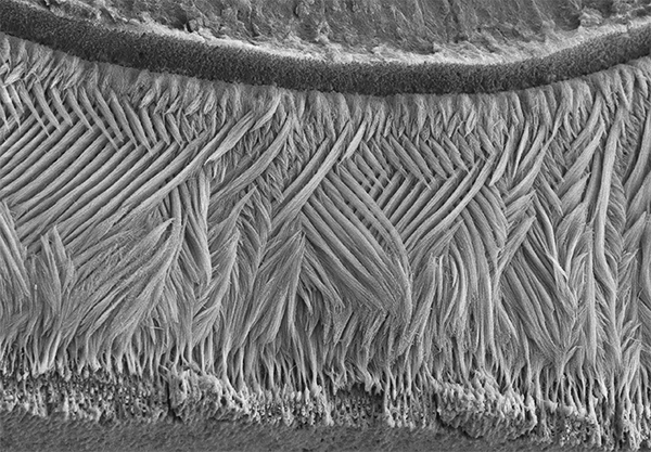
If you went out and asked folks what they’re seeing in this picture, most would probably guess an elegantly woven basket, or a soft, downy feather. But what this scanning electron micrograph actually shows isn’t at all soft: it is the hardest substance in the mammalian body—tooth enamel!
By IRP Staff Blogger
Wednesday, January 20, 2016
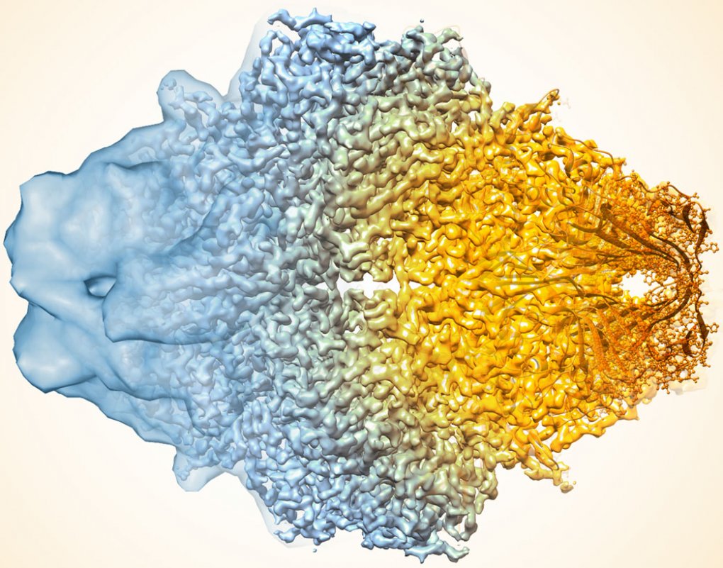
In the quest to find faster, better ways of mapping the structure of proteins and other key biological molecules, a growing number of researchers are turning to an innovative method that pushes the idea of a freeze frame to a whole new level: cryo-electron microscopy (cryo-EM).