Beat the Heat With Some NIH History
Measuring and Manipulating Temperature Is Key to IRP Research
Many parts of the country were hit by record-breaking heat this summer. Controlling temperature is not only important for staying healthy, but it's also crucial for many types of research. Grab your water bottle and sit down in a shady spot to take a look at some photos from the Office of NIH History & Stetten Museum on the theme of hot and cold.
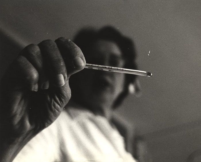
How do we measure temperature? One way is with a glass thermometer, as seen in this photo from the Jerry Hecht Collection. Hecht was an NIH photographer who visited many different laboratories, offices, institutes, and centers to take dramatic and artistic photos of people at work.
The mercury contained within the thermometer’s bulb expands as heat increases. This sends the mercury up the narrow tube, where we interpret it as temperature using numbers written along the side. Glass thermometers have been used since the first half of the 1700s — the time of Gabriel Fahrenheit and Andres Celsius. They each gave their name to a way to interpret the temperature.
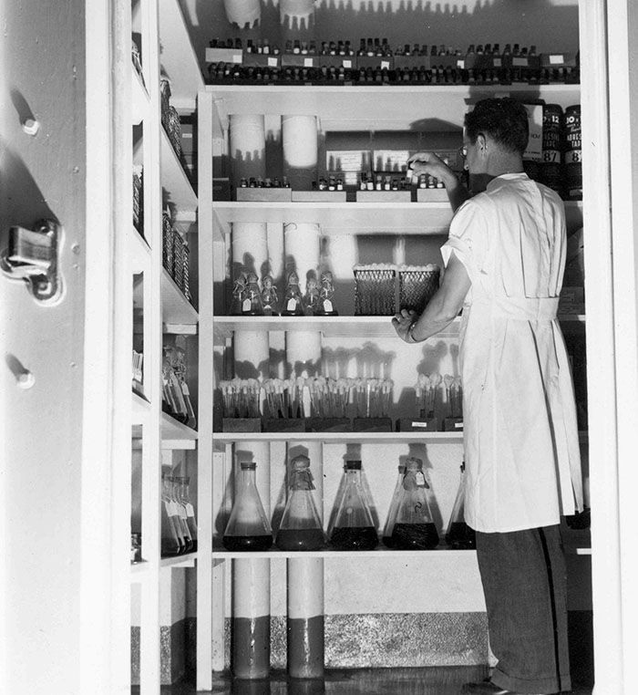
Need a break from the heat? Cold room storage, like this one pictured at the National Institute of Allergy and Infectious Diseases’ (NIAID) Rocky Mountain Labs campus in the 1940s, is kept at a chilly -176 degrees Fahrenheit. These frigid temperatures keep sensitive biological material from deteriorating. Researchers there were creating vaccines for Rocky Mountain spotted fever, typhus, and yellow fever. Many of these vials and flasks likely contained vaccine experiments and tick samples.
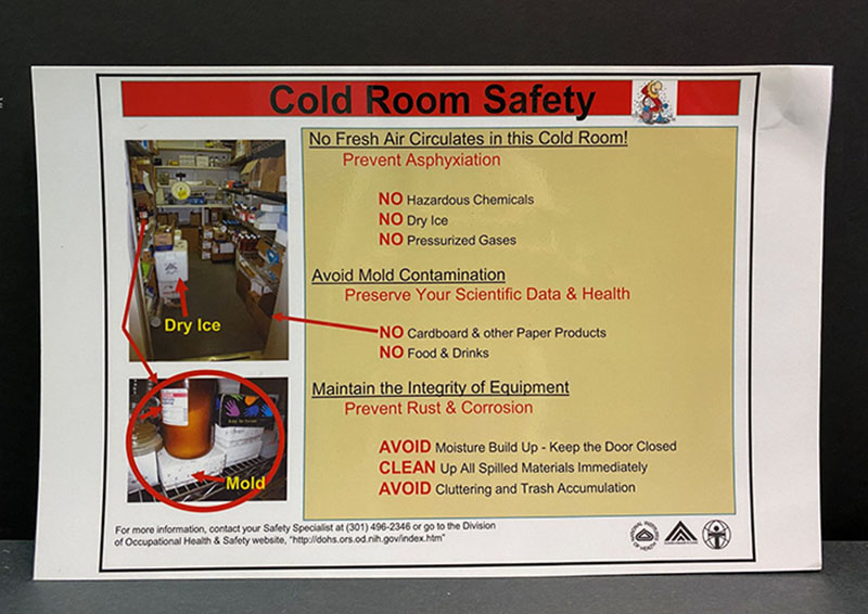
A cold room sounds nice and refreshing on a hot day, but at NIH, safety always comes first. This cold room safety sign was posted in Building 29A, which housed the FDA’s Center for Biologics Evaluation and Research. As this sign explains, these rooms are sealed and researchers have to be careful not to bring any contaminants inside. Mold, rust, and corrosion can still occur — even at -176 degrees Fahrenheit.
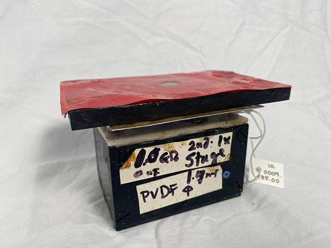
Sometimes researchers at NIH have to get creative. IRP investigator Ichiji Tasaki, M.D., couldn’t find a sensor sensitive enough to detect small, brief moments of heat in cells and tissues, so he decided to make one of his own. He used Piezo film, a flexible, lightweight plastic that can be molded into custom shapes and measures the amplitude, frequency, and direction of heat events. Using his device, he was able to find the exact location of heating within a cell. In 1999, he published the paper “Evidence for Phase Transition in Nerve Fibers, Cells, and Synapses,” where he praised this new machine for “detecting small and brief heat production in biological tissues.”
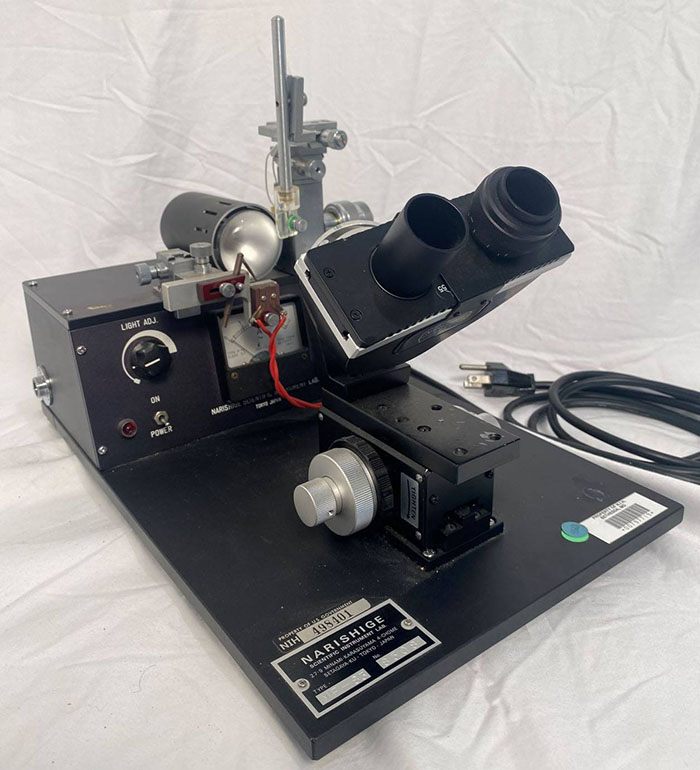
While blacksmiths have forges, scientists have microforges. Microforges like this one are akin to a small glass-blowing studio, using heat to melt, bend, and form pipette tips specifically suited to a researcher’s needs. This work happens on a very small scale and the eyepieces on the device allow for a microscopic view of the pipette tip.
This microforge was donated by Marshall Nirenberg, Ph.D., an IRP researcher who helped crack the genetic code. Dr. Nirenberg earned the 1968 Nobel Prize in Physiology and Medicine for his discovery.
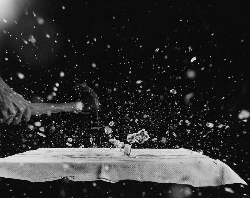
This photo gives new meaning to the phrase “breaking the ice.” The image is part of the Randolph A. Kennedy collection in the archives of the Office of NIH History and Stetten Museum. Kennedy worked at NIH in the 1950s and 60s in the Photographic Section of the Scientific Review Branch in the Division of Research Services. He started photography as a hobby when he was only 12 years old and living in Clifton Forge, Virginia. After high school, he followed his passion and attended the National School of Photography in Silver Spring, Maryland. In 1949, he was hired at NIH, where he took portraits and laboratory photos, processed film, made prints, and contributed heavily to the NIH Record. You can read more about him in a Spotlight article on page three of this 1955 edition of the Record.
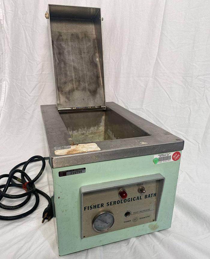
Is it getting hot in here? Serological baths like this one produce a slow, consistent application of heat for bacteria samples. When paired with the right media in which to grow the bacteria, they can speed up the growth process. You probably don’t want bacteria growing in your house, but scientists at NIH frequently need to grow large amounts of bacteria for their research.
This serological bath from 1978 was used by IRP investigator Alan Peterkofsky, Ph.D., head of the Macromolecules Section of the Laboratory of Biochemical Genetics at the National Heart, Lung, and Blood Institute (NHLBI). Over the course of his career, he sought to uncover how cyclic AMP, a biological messenger molecule, is regulated in bacteria. Cyclic AMP is used for communication within cells and is particularly important in regulating hormone function.
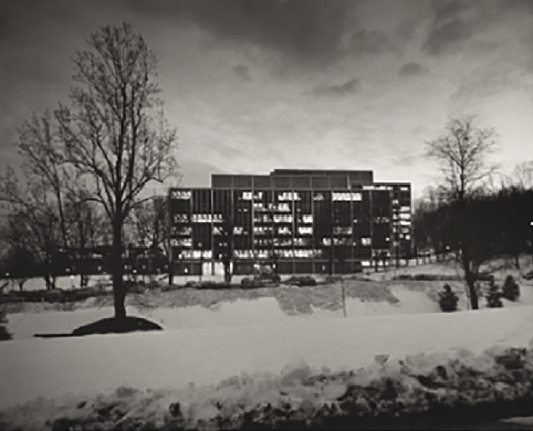
Are you looking forward to the colder months? After the heat of summer, this snowy photo of Building 31 on NIH’s Bethesda campus is looking very appealing. The photo is also from our Jerry Hecht collection. As we continue to contend with toasty outdoor temperatures, maybe bringing pictures like this one to mind will help us tough it out until fall rolls around.
Subscribe to our weekly newsletter to stay up-to-date on the latest breakthroughs in the NIH Intramural Research Program.
Related Blog Posts
This page was last updated on Monday, January 29, 2024
