Celebrating the Art of Science
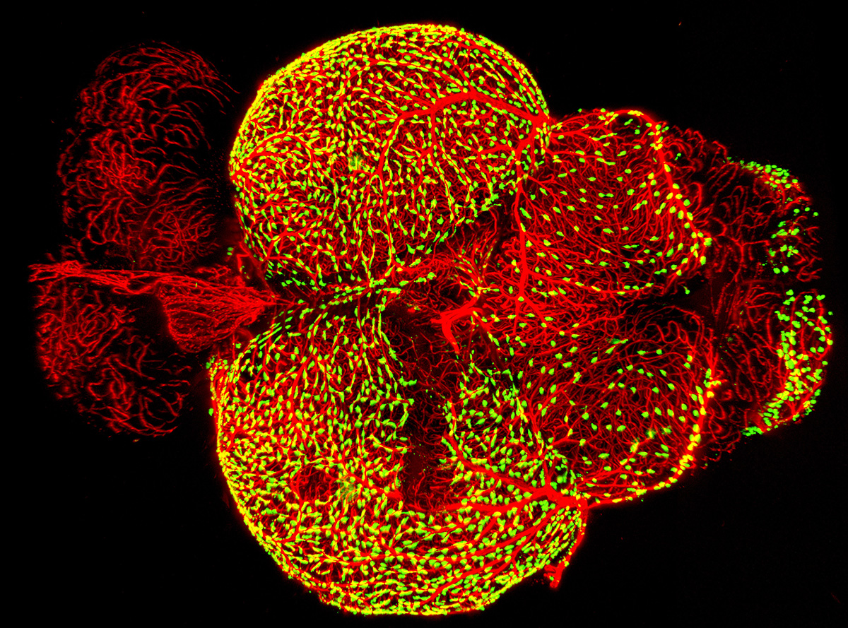
Blood Vessel Cells in a Zebrafish Brain

CREDIT: DAN CASTRANOVA, MARINA VENERO GALANTERNIK, AND BRANT WEINSTEIN (NICHD); TUYET NGUYEN (UNIVERSITY OF MARYLAND)
Scientists from the Eunice Kennedy Shriver National Institute of Child Health and Human Development (NICHD) produced this award-winning microscopy image that shows how fluorescent granular perithelial cells (FGPs, in green) are closely associated with blood vessels (red) that surround an adult zebrafish’s (Danio rerio) brain. The investigators recently discovered that FGPs, which are found in both zebrafish and mammals, are related to cells that form the lymphatic system and are thought to play a key role in maintaining the blood–brain barrier and clearing toxic substances from the brain (eLife 6:e24369, 2017; DOI:10.7554/eLife.24369). The image was selected as one of the winners of the 2017 BioArt competition held by the Federation of American Societies for Experimental Biology (FASEB).
Through its annual BioArt competition, FASEB aims to share the beauty and breadth of biological research with the public by celebrating the art of science. Contestants include investigators, contractors, and trainees with current or past research funding from a U.S. federal agency and members of FASEB societies. The 2017 competition featured 15 winning entries (12 images and three videos) that represented a wide range of biological research, from mercury toxicity to lung disease to hazardous-waste-storage safety. The NIH winning entry, entitled “Blood Vessel Cells in an Adult Zebrafish Brain,” was submitted by NICHD researchers—Marina Galanternik, Daniel Castranova, and Brant Weinstein—and Tuyet Nguyen (University of Maryland, College Park, Maryland).
“These images offer glimpses of the wide range of extraordinary scientific progress being made in labs across the country,” said FASEB President Thomas Baldwin. “They’re captivatingly beautiful.”
To see more winning images, go to the FASEB BioArt website.
This page was last updated on Friday, April 8, 2022
