Daniel Reich, M.D., Ph.D.: Imagining Brain Imaging
BY ANNE DAVIDSON, NICHD
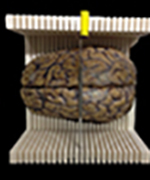
Humming along in the corner of Daniel Reich’s lab is a small scientific instrument that you’d expect to see at a tech company or in a design studio. It’s a 3-D printer busily making a customized cutting box that can hold a brain extracted at an autopsy. The indentation at the bottom of the box cradles the brain while the comb-like projections along the sides are used as guides for slicing the brain into thin sections that can be compared with magnetic resonance imaging (MRI) scans.

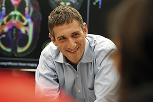
NINDS Senior Investigator Daniel Reich uses advanced MRI techniques to advance scientific knowledge about multiple sclerosis.
Reich, a neuroradiologist and senior investigator in the National Institute of Neurological Disorders and Stroke (NINDS), and his group developed the 3-D-printed cutting boxes as part of their research on multiple sclerosis (MS), a chronic neuroinflammatory autoimmune disorder of the central nervous system. MS affects some 400,000 Americans and about 2.5 million people worldwide and appears most often when people are between 20 and 40 years old and more commonly among women.
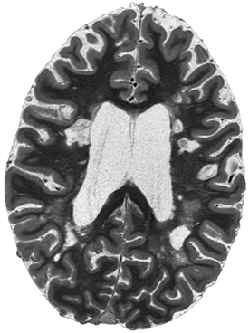
In MS, the immune system damages or destroys the myelin (which envelops and insulates the nerves) and causes plaques, or lesions, to form. The lesions show up as white spots on MRI scans of the brain. But the severity of the disease cannot be determined by scans alone. Even when symptoms—such as muscle weakness in the hands and legs, impaired vision, clumsiness, and other problems—worsen, MRI scans may not reflect an increase in the size or number of lesions. Conversely, new lesions that are visible on an MRI scan may not be accompanied by new symptoms. Such inconsistencies make MS a complicated disease to characterize and treat.

CREDIT: DANIEL REICH, NINDS
MS lesions show up as white spots in an MRI scan of the brain.
One of the ways that Reich is getting a better understanding of the pathological basis of MS is to do MRI-guided histopathology in which slices of postmortem brain tissue are compared with an MRI scan of the whole brain. That’s where the customized 3-D-printed cutting boxes come in. In the past, it was difficult to obtain a correlation between MRI images and standard pathological sections of a postmortem brain. Tiny lesions that were visible on an MRI scan could be hard to find in actual brain tissue because the sections tended to be relatively thick and the slice faces weren’t always parallel. But the 3-D printer can be programmed to create a mold to fit each brain exactly and have guides that ensured precision slicing. The use of the 3-D-printed cutting boxes can improve the speed, quality, and accuracy of finding—in brain tissue—MRI-identified small lesions that occur in MS and other brain diseases. Now this 3-D technique routinely helps Reich’s lab study the use of MRI as a biomarker for the progression of MS. (J Neuropathol Exp Neurol 73:780–788, 2014; DOI:10.1097/NEN.0000000000000096). For a how-to video of the protocol, see the Journal of Visualized Experiments (JoVE, (118), 2016; http://doi.org/10.3791/54780)

CREDIT: DANIEL REICH, NINDS
Reich’s lab developed a technique for using a 3-D printer to make customized cutting boxes. Shown: a cutting box cradles a postmortem brain; the guides on the sides of the box enable the researchers to cut the brain into thin, precise slices.
Reich also uses MRI imaging to study the progression of MS in live patients (he works with the NINDS Neuroimmunology Clinic and its chief, Irene Cortese). Most MRIs in ordinary medical settings operate at either 1.5 and 3 tesla in strength, but some MRIs used in research are much more powerful. The higher the strength, the stronger the MRI signal and the better the image quality. Reich uses a 7-tesla MRI machine to get very detailed, high-resolution images that have provided new insights into the pathological changes of newly forming lesions. He’s found that lesions evolve through different stages: centrifugal, where inflammation derives from a small blood vessel in the center of the lesion; and centripetal, where blood-derived inflammatory cells and molecules start from the rim and flow inward (Nature Reviews Neurology 12:358–368, 2016; http://doi.org/10.1038/nrneurol.2016.59). Reich has also learned to identify a group of lesions that are chronically inflamed and a possible target for new drugs.
Building on a Long History of MS Research
Reich’s research is built on decades of MRI data collected at NIH. In the 1980s, NINDS scientist Henry McFarland pioneered the use of MRI to study MS. He was responsible for hiring Reich in 2009 and then mentoring him. In the 1990s and early 2000s, McFarland and Joseph Frank (Clinical Center) used marmosets as a primate model for MS research. Reich and NINDS senior investigators Steven Jacobson and Afonso Silva re-established the model at NIH in 2010.
Usually, scientists who want to study MS and other brain inflammatory diseases use a mouse model called experimental autoimmune encephalomyelitis (EAE). But the pathology of EAE in rodents is markedly different than that of MS in humans. When given EAE, marmosets, however develop lesions and symptoms similar to human MS. Marmoset brains are smoother and have fewer ridges than human brains and are in some ways easier to image. And breeding the animals is easy: the females give birth to nonidentical twins or triplets per delivery and can deliver twice a year. Marmoset MS research is something very few other labs have the facilities to undertake. Using marmosets, the Reich lab has been able to noninvasively monitor pre-lesional changes in MS and has shown that blood–brain barrier permeability increases about four weeks before demyelination. The lab is now working to develop the model further to study chronic MS-type lesions and how they can be repaired.
Deciding to Become a Scientist
Reich didn’t always know he was destined to become a biomedical researcher. He was drawn to politics, too. Even after a summer research fellowship at NINDS in 1990, he went on to found a political magazine at Yale University (New Haven, Connecticut) where he was an undergraduate majoring in mathematics and physics. But late one night in 1992, when he was working as a summer student in a lab at Rockefeller University (New York), he decided to become a scientist. As he was analyzing single-neuron data recorded from the thalamus of cats, he observed precise firing patterns in response to slowly and smoothly varying visual stimuli—something no one had seen before. That feeling of discovery got him hooked, and the finding was the subject of his first scientific paper. But this newfound vocational dream didn’t stop Reich (who has varied interests—and recently read War and Peace for the third time) from taking off for a semester during his senior year at Yale to work on the Bill Clinton-Al Gore presidential campaign in Little Rock, Arkansas.
Still, Reich’s passion for scientific research, combined with the spirit of exploration, characterizes his career. He is grateful to his NIH mentors—especially now-scientist-emeritus Henry McFarland, with whom he keeps in close touch—who gave him latitude to try new things and learn from his own mistakes. Reich, who trains young investigators now, advises them to not be afraid of the new—whether it be new opportunities, new techniques, new collaborations, or just new, surprising data.
DANIEL S. REICH, M.D., PH.D., NINDS
Senior Investigator and Chief, Translational Neuroradiology Section, Division of Neuroimmunology and Neurovirology, National Institute of Neurological Disorders and Stroke
Born: New Haven, Conn.
Grew up: Chevy Chase, Md.
Research Interests: Developing new magnetic-resonance imaging methods to investigate the origin of disability in multiple sclerosis and related disorders and applying those methods to patient care and to clinical trials of new drugs.
Education: Yale University, New Haven (B.S. in mathematics and physics); Weill Medical College of Cornell University and The Rockefeller University, New York (M.D. and Ph.D. in visual neurophysiology)
Training: Summer undergraduate internship at NINDS; residency in neurology/diagnostic radiology/neuroradiology at Johns Hopkins Hospital (Baltimore)
Came to NIH: In 1987 as a high school student to do a project (on a condition called pseudothrombocytopenia) in NHLBI; returned in 1990 for a summer research fellowship at NINDS; returned again in 2009 as chief of NINDS’s Translational Neuroradiology Unit; became senior investigator in 2016
Outside interests: Playing chamber music (violin and viola); traveling; running; biking; reading
Little Known Fact: Trained himself to listen to podcasts and audiobooks at twice the normal speed. “One thing that makes me very happy is to be listening to an audiobook while running and then to find myself, unexpectedly, in the very place that’s being described,” he said. “That happened to me once in Chelsea, London, when I was listening to Vanity Fair [by William Makepeace Thackeray], and it happened again this summer in Cape Cod while listening to Melvyn Bragg’s fascinating The Adventure of English: The Biography of a Language.”
Interesting Facts About His Family: Reich lives with his wife, a poet and teacher of literature and writing at the Maryland Institute College of Art (Baltimore), and daughter and son,who are middle school students at DC International Public Charter School. His sister is a senior lecturer in Slavic studies at the University of Cambridge (Cambridge, England), where she reads and writes about late Soviet dissident literature. His brother is a professor of genetics at Harvard Medical School (Boston) and is deeply involved in the Ancient DNA Revolution. His mother is the author of five satirical but serious novels that deal with the interactions among family, religion, politics, and the modern human condition. His father, a professor of psychiatry and international affairs at George Washington University (Washington, D.C.), is the former director of the United States Holocaust Memorial Museum (Washington, D.C.) and spent many years at the National Institute of Mental Health studying the use and abuse of psychiatry in the Soviet Union.
Website: https://irp.nih.gov/pi/daniel-reich
This page was last updated on Saturday, January 18, 2025
