Microscopy as Masterpiece
A Beautiful Way to Study Neurons

CREDIT: MARK STOPFER, NICHD
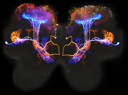
This composite image shows two neurons in the locust brain that process olfactory information.
Perhaps only a scientist can find the beauty within a locust brain. An image—looking like mirrored, psychedelic-colored mushrooms—captured by Mark Stopfer—reveals the intricacies of neurons in the brain of the Schistocerca Americana locust. The image is one of several included in the “Microscopy as Masterpiece” digital exhibit at the Strathmore arts center in North Bethesda, Maryland. NIH and the Strathmore partnered to develop the exhibit, which complements the “Arts and the Brain” lecture series and features beautiful brain microscopy images and videos created by NIH-supported researchers.
Stopfer, a senior investigator at the National Institute of Child Health and Human Development, uses simple animal models such as locusts to study basic questions in neuroscience. In particular, he is identifying and analyzing the properties of neurons that process information about odors. He places an intracellular electrode into a locust neuron to record responses when odors are puffed onto the animal’s antennae. The neurons respond with vigorous spiking when odors are presented; when the odor changes, the spiking patterns change, too.
Stopfer had the following to say about how the image was produced:
“Our goal in this project was to identify and analyze the properties of neurons that process information about odors. The image is a composite of several micrographs illustrating two kinds of olfactory neurons in the locust [(Schistocerca americana)] brain. In the background, in gray, is an outline of the locust brain. Superimposed on that, in a whole assortment of colors, is a confocal microscope image of the ‘mushroom bodies,’ brain areas that process sensory stimuli and form memories. The image was made by injecting large amounts of dye into the mushroom bodies and then imaging them such that parts closer to the brain surface are more red and parts deeper in the brain are more blue.
“Finally, the image includes two olfactory neurons. In the intact animal, each neuron was studied by placing an intracellular electrode into it to record its responses when odors were puffed onto the animal’s antennae. (Both neurons respond with vigorous spiking when odors are presented; the spiking patterns change when the odor is changed.) Then, dye was injected into the neuron, and later the neuron was imaged on a confocal microscope. One cell here is colored blue and one orange. These colors were selected artificially in Photoshop when the composite image was assembled. Overall, the image shows the locations, enormous size, and complex branching patterns of neurons that process olfactory information.”
Arts and the Brain Lecture Series
Strathmore’s “Arts and the Brain” lecture series engages teachers, scholars, and artists working at the intersection of arts and health to present innovative, practical strategies for harnessing the arts to alleviate suffering and strengthen vitality. The final lecture, titled “Medical Avatar,” will take place on Thursday, June 1, at 7:30 p.m., in the Mansion at Strathmore. For information, go to https://www.strathmore.org/education/programs-for-adults/arts-the-brain-package. Tickets cost $25 for each lecture, but you can view the “Microscopy as Masterpiece” exhibit for free.

CREDIT: JEAN LIVET, TAMILY A. WEISSMAN, JEFF W. LICHTMAN, HARVARD UNIVERSITY
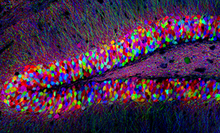
Random mixing of fluorescent dyes created this psychedelic slice of a mouse brain. Known as “brainbow,” the technique allows scientists to distinguish nearby cells by color and has helped advance the NIH Human Connectome Project.

CREDIT: KEUNYOUNG KIM, WONKYU JU AND MARK ELLISMAN, NATIONAL CENTER FOR MICROSCOPY AND IMAGING RESEARCH, DEPARTMENT OF OPHTHALMOLOGY, UNIVERSITY OF CALIFORNIA, SAN DIEGO
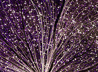
The pinpricks of light in this starry night sky are actually green fluorescent protein, marking specific cells in a mouse retina. This detailed image was made using large-scale mosaic confocal microscopy, a technique that, like Google Earth, computationally stitches together many small, high-resolution images.
This page was last updated on Monday, April 11, 2022
