Research advances from the National Institutes of Health (NIH) Intramural Research Program (IRP) often make headlines. Read the news releases that describe our most recent findings:
Findings from a phase 2 clinical trial show that the drug selumetinib improves outcomes for children with the genetic disorder neurofibromatosis type 1 (NF1). In the trial, selumetinib shrank the inoperable tumors that develop with NF1 called plexiform neurofibromas, and children experienced reduced pain, improved function, and better overall quality of life after receiving the treatment.
The trial was led by intramural researchers in the Center for Cancer Research (CCR) at the National Cancer Institute (NCI), part of the National Institutes of Health. Results of the trial were published March 18, 2020, in the New England Journal of Medicine.
“Until now, no effective medical therapies have existed for children with NF1 and plexiform neurofibromas, and it’s been a long journey to find a drug that can help them,” said Brigitte Widemann, M.D., lead author of the study, and chief of CCR’s Pediatric Oncology Branch, which developed and coordinated the trial. “While this is not yet a cure, this treatment is shrinking tumors and it’s making children feel better and have a better quality of life.”
Eva Dombi, M.D., Trish Whitcomb, R.N., Brigitte Widemann, M.D., Andrea Gross, M.D., and Andrea Baldwin, C.R.N.P., of the Pediatric Oncology Branch at NCI.
The virus that causes coronavirus disease 2019 (COVID-19) is stable for several hours to days in aerosols and on surfaces, according to a new study from National Institutes of Health, CDC, UCLA and Princeton University scientists in The New England Journal of Medicine. The scientists found that severe acute respiratory syndrome coronavirus 2 (SARS-CoV-2) was detectable in aerosols for up to three hours, up to four hours on copper, up to 24 hours on cardboard and up to two to three days on plastic and stainless steel. The results provide key information about the stability of SARS-CoV-2, which causes COVID-19 disease, and suggests that people may acquire the virus through the air and after touching contaminated objects. The study information was widely shared during the past two weeks after the researchers placed the contents on a preprint server to quickly share their data with colleagues.
The NIH scientists, from the National Institute of Allergy and Infectious Diseases’ Montana facility at Rocky Mountain Laboratories, compared how the environment affects SARS-CoV-2 and SARS-CoV-1, which causes SARS. SARS-CoV-1, like its successor now circulating across the globe, emerged from China and infected more than 8,000 people in 2002 and 2003. SARS-CoV-1 was eradicated by intensive contact tracing and case isolation measures and no cases have been detected since 2004. SARS-CoV-1 is the human coronavirus most closely related to SARS-CoV-2. In the stability study the two viruses behaved similarly, which unfortunately fails to explain why COVID-19 has become a much larger outbreak.
The NIH study attempted to mimic virus being deposited from an infected person onto everyday surfaces in a household or hospital setting, such as through coughing or touching objects. The scientists then investigated how long the virus remained infectious on these surfaces.
This scanning electron microscope image shows SARS-CoV-2 (yellow) — also known as 2019-nCoV, the virus that causes COVID-19 — isolated from a patient in the U.S., emerging from the surface of cells (blue/pink) cultured in the lab.
Finding may lead to novel therapeutic target for blinding disease
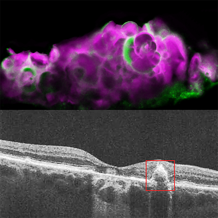
A protein that normally deposits mineralized calcium in tooth enamel may also be responsible for calcium deposits in the back of the eye in people with dry age-related macular degeneration (AMD), according to a study from researchers at the National Eye Institute (NEI). This protein, amelotin, may turn out to be a therapeutic target for the blinding disease. The findings were published in the journal Translational Research. NEI is part of the National Institutes of Health.
“Using a simple cell culture model of retinal pigment epithelial cells, we were able to show that amelotin gets turned on by a certain kind of stress and causes formation of a particular kind of calcium deposit also seen in bones and teeth. When we looked in human donor eyes with dry AMD, we saw the same thing,” said Graeme Wistow, Ph.D., chief of the NEI Section on Molecular Structure and Functional Genomics, and senior author of the study.
There are two forms of AMD — wet and dry. While there are treatments that can slow the progression of wet AMD, there are currently no treatments for dry AMD, also called geographic atrophy. In dry AMD, deposits of cholesterol, lipids, proteins, and minerals accumulate at the back of the eye. Some of these deposits are called soft drusen and have a specific composition, different from deposits found in wet AMD. Drusen form under the retinal pigment epithelium (RPE), a layer of cells that transports nutrients from the blood vessels below to support the light-sensing photoreceptors of the retina above them. As the drusen develop, the RPE and eventually the photoreceptors die, leading to blindness. The photoreceptors cannot grow back, so the blindness is permanent.
Top: HAP spherules (pink) and amelotin protein (green) in soft drusen from eye with dry AMD. Bottom: OCT image of eye with dry AMD, showing soft drusen beneath the retinal pigment epithelium.
Disabling key enzymes overcomes previous limitations to blocking angiogenesis, may inform cancer treatment strategies
Scientists at the National Institutes of Health and other institutions have devised a new strategy to stop tumors from developing the new blood vessels they need to grow. Once thought to be extremely promising for the treatment of cancer, blocking molecules that stimulate new blood vessel growth (angiogenesis) has proven ineffective because tumor cells respond by producing more stimulatory molecules. The new strategy involves disabling key enzymes that replenish the molecule that cells need for the reactions that sustain new vessel growth. The research team was led by Brant M. Weinstein, Ph.D., chief of the Section on Vertebrate Organogenesis at NIH’s Eunice Kennedy Shriver National Institute of Child Health and Human Development (NICHD). The study appears in Nature Communications.
Among the angiogenesis factors that stimulate new vessel growth is vascular endothelial growth factor (VEGF), which binds to a receptor on cell surfaces. This binding sets off a sequence of chemical reactions inside the cells lining the inside of blood vessels, culminating in new vessel growth. Previous attempts have sought to prevent this binding by targeting VEGF with antibodies or drugs, or by blocking the receptor so VEGF can’t bind to it. However, tumors respond by producing more VEGF, overwhelming such efforts.
After binding occurs, an enzyme that converts the compound phosphatidylinositol-(4,5)-bisphosphate (PIP2) into inositol triphosphate, which is needed for the reactions that fuel new blood vessel growth, and diacylglycerol (DAG). Through a series of enzyme-assisted steps, DAG is converted back into PIP2, allowing it to be recycled, as needed.
The researchers showed that they could stop angiogenesis by blocking any of the enzymes in this PIP2 recycling series. They first halted angiogenesis in human cell cultures and zebrafish embryos by disabling the genes for one or more of the enzymes. They then targeted tumors in mice with drugs that block the recycling enzymes. Compared to normal mice, the treated mice had less tumor and tumor blood vessel growth. Moreover, adding more VEGF depleted any remaining PIP2, further reducing blood vessel growth.
New strategy could be applied to other infectious diseases
A new approach to direct the body to make a specific antibody against HIV led to sustained production of that antibody for more than a year among participants in a National Institutes of Health clinical trial. This drug-delivery technology uses a harmless virus to deliver an antibody gene into human cells, enabling the body to generate the antibody over an extended time. With further development, such a strategy could be applied to prevent and treat a wide variety of infectious diseases, according to the study investigators.
Researchers from NIH’s National Institute of Allergy and Infectious Diseases (NIAID) reported the findings on March 9 in an oral presentation at the 2020 Conference on Retroviruses and Opportunistic Infections (CROI).
Antibodies are immune system proteins that help prevent or clear infections. Traditional vaccines induce the immune system to generate protective antibodies. Another approach to preventing infections is to deliver monoclonal antibodies — preparations of a specific antibody designed to bind to a single target — directly into people. Monoclonal antibodies also are used therapeutically, with many already approved for treating cancer, autoimmune diseases and other conditions and others being evaluated for treatment of infectious diseases, such as Ebola virus disease.
Administering proteins to people requires periodic injections or infusions to retain protective or therapeutic levels, which can be challenging, particularly in resource-limited settings. Delivery of antibody genes using a virus as a carrier, or vector, offers a potential alternative.
The new drug-delivery technology uses a harmless virus to deliver an antibody gene into human cells.
NIH study suggests our brains use distinct firing patterns to store and replay memories
In a study of epilepsy patients, researchers at the National Institutes of Health monitored the electrical activity of thousands of individual brain cells, called neurons, as patients took memory tests. They found that the firing patterns of the cells that occurred when patients learned a word pair were replayed fractions of a second before they successfully remembered the pair. The study was part of an NIH Clinical Center trial for patients with drug-resistant epilepsy whose seizures cannot be controlled with drugs.
“Memory plays a crucial role in our lives. Just as musical notes are recorded as grooves on a record, it appears that our brains store memories in neural firing patterns that can be replayed over and over again,” said Kareem Zaghloul, M.D., Ph.D., a neurosurgeon-researcher at the NIH’s National Institute of Neurological Disorders and Stroke (NINDS) and senior author of the study published in Science.
Dr. Zaghloul’s team has been recording electrical currents of drug-resistant epilepsy patients temporarily living with surgically implanted electrodes designed to monitor brain activity in the hopes of identifying the source of a patient’s seizures. This period also provides an opportunity to study neural activity during memory. In this study, his team examined the activity used to store memories of our past experiences, which scientists call episodic memories.
IRP researchers found that our brains may store memories in neuronal firing patterns that are replayed fractions of a second before remembering.
Researchers at the National Institutes of Health and the University of Wisconsin have demonstrated that using artificial intelligence to analyze CT scans can produce more accurate risk assessment for major cardiovascular events than current, standard methods such as the Framingham risk score (FRS) and body-mass index (BMI).
More than 80 million body CT scans are performed every year in the U.S. alone, but valuable prognostic information on body composition is typically overlooked. In this study, for example, abdominal scans done for routine colorectal cancer screening revealed important information about heart-related risks – when AI was used to analyze the images.
The study compared the ability of automated CT-based body composition biomarkers derived from image-processing algorithms to predict major cardiovascular events and overall survival against routinely used clinical parameters. The investigators found that the CT-based measures were more accurate than FRS and BMI in predicting downstream adverse events including death or myocardial infarction, cerebrovascular accident, or congestive heart failure. The results appeared in The Lancet Digital Health.
“We found that automated measures provided more accurate risk assessments than established clinical biomarkers,” said Ronald M. Summers, M.D., Ph.D., of the NIH Clinical Center and senior author of the study. “This demonstrates the potential of an approach that uses AI to tap into the biometric data embedded in all such scans performed for a wide range of other indications and derive information that can help people better understand their overall health and risks of serious adverse events.”
Improved diagnosis could reduce the risk of both overtreatment and undertreatment of the disease
A method of testing for prostate cancer developed at the National Cancer Institute (NCI) leads to more accurate diagnosis and prediction of the course of the disease, according to a large study. This method, which combines systematic biopsy, the current primary diagnostic approach, with MRI-targeted biopsy, is poised to greatly improve prostate cancer diagnosis, thereby reducing the risk of both overtreatment and undertreatment of the disease. NCI is part of the National Institutes of Health.
The findings were published March 5, 2020, in the New England Journal of Medicine. The study was conducted at the NIH Clinical Center in Bethesda, Maryland.
“Prostate cancer has been one of the only solid tumors diagnosed by performing systematic biopsies ‘blind’ to the cancer’s location. For decades this has led to the overdiagnosis and subsequent unnecessary treatment of non-lethal cancers, as well as to missing aggressive high-grade cancers and their opportunity for cure,” said Peter Pinto, M.D., of the Urologic Oncology Branch in NCI’s Center for Cancer Research and senior author of the study. “With the addition of MRI-targeted biopsy to systematic biopsy, we can now identify the most lethal cancers within the prostate earlier, providing patients the potential for better treatment before the cancers spread.”
A 3-D map of the prostate using combined MRI-targeted and systematic biopsies. Using both types of biopsy greatly improved prostate cancer diagnosis in a new study.
NIH-funded project in mice provides insights into why nerves fail to regrow following injury
When the spinal cord is injured, the damaged nerve fibers — called axons — are normally incapable of regrowth, leading to permanent loss of function. Considerable research has been done to find ways to promote the regeneration of axons following injury. Results of a study performed in mice and published in Cell Metabolism suggests that increasing energy supply within these injured spinal cord nerves could help promote axon regrowth and restore some motor functions. The study was a collaboration between the National Institutes of Health and the Indiana University School of Medicine in Indianapolis.
“We are the first to show that spinal cord injury results in an energy crisis that is intrinsically linked to the limited ability of damaged axons to regenerate,” said Zu-Hang Sheng, Ph.D., senior principal investigator at the NIH’s National Institute of Neurological Disorders and Stroke (NINDS) and a co-senior author of the study.
Like gasoline for a car engine, the cells of the body use a chemical compound called adenosine triphosphate (ATP) for fuel. Much of this ATP is made by cellular power plants called mitochondria. In spinal cord nerves, mitochondria can be found along the axons. When axons are injured, the nearby mitochondria are often damaged as well, impairing ATP production in injured nerves.
“Nerve repair requires a significant amount of energy,” said Dr. Sheng. “Our hypothesis is that damage to mitochondria following injury severely limits the available ATP, and this energy crisis is what prevents the regrowth and repair of injured axons.”
After injury (see damage at the center of the image), nerve fibers (in red) regrow past the injury (right) when energy levels in the tissue are increased.
Findings suggest drugs targeting immune cells may help treat deadly disease mainly affecting children
Researchers at the National Institutes of Health found evidence that specific immune cells may play a key role in the devastating effects of cerebral malaria, a severe form of malaria that mainly affects young children. The results, published in the Journal of Clinical Investigation, suggest that drugs targeting T cells may be effective in treating the disease. The study was supported by the NIH Intramural Research Program.
“This is the first study showing that T cells target blood vessels in brains of children with cerebral malaria,” said Dorian McGavern, Ph.D., chief of the Viral Immunology and Intravital Imaging Section at the NIH’s National Institute of Neurological Disorders and Stroke (NINDS) who co-directed the study with Susan Pierce, Ph.D., chief of the Laboratory of Immunogenetics at the National Institute of Allergy and Infectious Diseases (NIAID). “These findings build a bridge between mouse and human cerebral malaria studies by implicating T cells in the development of disease pathology in children. It is well established that T cells cause the brain vasculature injury associated with cerebral malaria in mice, but this was not known in humans.”
More than 200 million people worldwide are infected annually with mosquito-borne parasites that cause malaria. In a subset of those patients, mainly young children, the parasites accumulate in brain blood vessels causing cerebral malaria, which leads to increased brain pressure from swelling. Even with available treatment, cerebral malaria still kills up to 25% of those affected resulting in nearly 400,000 deaths annually. Children who survive the infection will often have long-lasting neurological problems such as cognitive impairment.
Specific immune cells accumulate within brain blood vessels of people affected by cerebral malaria. This finding suggests a new treatment strategy for the disease.