Colleagues: Recently Tenured
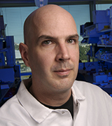
KEVIN M. BROWN, PH.D., NCI-DCEG
Senior Investigator, Laboratory of Translational Genomics, Division of Cancer Epidemiology and Genetics, National Cancer Institute

Education: University of Virginia, Charlottesville, Virginia (B.A. in biology); George Washington University, Washington, D.C. (Ph.D. in genetics)
Training: Postdoctoral fellow, Genetic Basis for Human Disease Division, Translational Genomics Research Institute (TGen, Phoenix)
Before coming to NIH: Investigator, Integrative Cancer Genomics Division, TGen; adjunct professor of Genetic Epidemiology, Mayo Clinic Cancer Center (Scottsdale, Arizona); adjunct research assistant professor, Basic Medical Sciences, University of Arizona College of Medicine (Phoenix); adjunct professor, Molecular and Cellular Biology, Arizona State University (Tempe, Arizona)
Came to NIH: In 2010
Selected professional activities: Workshop faculty, American Association for Cancer Research’s Integrative Molecular Epidemiology Workshop; member, American Cancer Society’s Tumor Biology and Genomics Study Section
Outside interests: Mountain biking; barbequing
Website: https://irp.nih.gov/pi/kevin-brown
Research interests: My research focuses on the genetic underpinnings of melanoma susceptibility. My lab is identifying the genetic contributions and functional pathways associated with melanoma risk. Although early-stage melanoma is largely curable via surgical resection, outcomes for a significant proportion of late-stage melanoma patients remain poor despite considerable recent progress.
We are trying to gain a better understanding of the genetic factors contributing to melanoma risk so we can facilitate prevention and/or early-detection efforts in at-risk individuals. Also, we are actively involved in ongoing melanoma genome-wide association studies (GWAS) as members of the International Melanoma Genetics Consortium (GenoMEL) and the Melanoma Meta-analysis Consortium. We also work with GenoMEL member groups to use whole-genome and whole-exome sequencing approaches to better understand the genetics of melanoma susceptibility in melanoma-prone families. Lastly, we are also actively involved in ongoing renal-cell cancer GWAS at NCI, as well as meta-analyses with international teams working in the same area.
Beyond identification of novel risk genes and loci, a significant proportion of my research program focuses on functional characterization of cancer-susceptibility loci identified via GWAS. Although significant strides have been made in recent years toward cataloging loci involved in cancer risk, research into how these variants influence risk lags far behind. We are trying to better understand the molecular consequences of risk variants on gene regulation and function as well as the phenotypic effects of allele-specific gene functions. We perform fine-mapping of risk-associated regions, targeted and genome-wide expression analysis, gene-regulation and epigenetic work, and bioinformatic analysis of large genomic datasets.
Our current projects include a large-scale study of the influences of germline genetic variation on both gene regulation and cellular phenotypes in melanocytes and melanoma cells; functional genomic screens to identify genes that mediate phenotypes associated with early-stage tumor progression; large-scale reporter screens to identify risk-associated genetic variants conferring allele-specific gene regulatory potential; and directed analyses applied to specific susceptibility regions and genes.
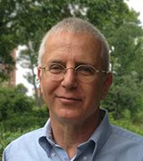
AHMED M. GHARIB, M.D., NIDDK
Senior Investigator; Head, Biomedical and Metabolic Imaging Branch, National Institute of Diabetes and Digestive and Kidney Diseases

Education: University of Alexandria, Faculty of Medicine, Alexandria, Egypt (M.D.)
Training: Internship at Alexandria University Hospital, Egypt, and in internal medicine at University of Washington (Seattle); residency in nuclear medicine and nuclear cardiology at University of Washington; residency in diagnostic radiology at University of Louisville (Louisville, Ky.); fellow/instructor in cross-sectional imaging at Johns Hopkins Hospital (Baltimore); and fellow in NIH’s Imaging Science Training Program
Came to NIH: In 2003 for training; 2004–2005 served as staff clinician in the NIH Clinical Center’s Diagnostic Radiology Department, then in NHLBI’s Integrated Cardiovascular Imaging Laboratory, and later in NIDDK’s Biomedical and Metabolic Imaging Branch; in 2011 became tenure-track investigator
Selected professional activities: Member, research committee, North American Society of Cardiovascular Imaging; member, Cardiovascular Radiology and Intervention Communications Committee, American Heart Association; member, NIH Radiation Safety Committee
Outside interests: Enjoys motor sports including cars, motorcycles, and mechanics; spending time with his family
Research interests: My research involves developing new imaging techniques to detect organ damage associated with metabolic and immunological disorders. In 2013, I received the NIH Director’s Award for the “development of cutting edge, non-invasive imaging technologies permitting drastically improved resolution and disease detection while reducing the risk to patients.”
NIDDK’s Biomedical and Metabolic Imaging Branch (BMIB)—which is made up of a multidisciplinary team of biomedical engineers, spectroscopy experts, and biologists—is using new chemical spectroscopic techniques to view organs (such as the liver, heart, and muscles) and improved imaging methodologies (that are faster and are at a higher resolution), and high-contrast techniques without the use of contrast agents.
We applied both computed tomography (CT) and magnetic-resonance imaging (MRI) for coronary imaging in the same patients. This approach allowed for identifying coronary abnormalities in people with HIV, Job syndrome, and Cushing syndrome. We also developed a new technique to image the coronary vessel wall—to characterize atherosclerotic coronary-artery disease—using high-magnetic-field MR scanners without the need for ionizing radiation. A patent application is pending for this new method, which could potentially be used to characterize and study coronary artery disease and its response to various lipid-lowering and anti-inflammatory therapies. These imaging techniques have also provided data for mathematical models that identify sites of maximum strain on the coronary arteries to predict points of potential atherosclerotic plaque accumulation and possible plaque vulnerability.
As part of the NIH obesity initiative, we have integrated the use of a large-bore high-magnetic-field MR scanner with a metabolic unit to characterize metabolic activity in people with a wide range of body-mass indices. An improvement and technical advancement in MR spectroscopy, developed by the BMIB team, has also been applied to measure fat and certain metabolites in the heart, liver, pancreas, and muscles and to correlate measurements with metabolic activity. We are also using imaging techniques that will allow for early detection and simultaneous quantification of liver fibrosis and inflammation in patients.
EYTAN RUPPIN, M.D., PH.D, NCI-CCR
Senior Investigator, Chief, Cancer Data Science Laboratory, Center for Cancer Research, National Cancer Institute

Education: Tel Aviv University, Tel Aviv, Israel (M.D.; M.Sc. and Ph.D. in computer science)
Training: Internship in medicine at Rabin Medical Center (Petah Tikva, Israel); residency in medicine and psychiatry at Ramat-Chen Psychiatric Center (Tel Aviv); postdoctoral training in computer science at the University of Maryland at College Park (College Park, Md.)
Before coming to NIH: Professor of computer science and medicine, Tel Aviv University; professor and director of Center for Bioinformatics and Computational Biology, University of Maryland at College Park
Came to NIH: In January 2018
Outside interests: Playing bridge; hiking
Website: https://ccr.cancer.gov/cancer-data-science-laboratory/eytan-ruppin
Research interests: I am helping to develop and harness computational systems–biology approaches for the multi-omics analysis (genome, proteome, transcriptome, epigenome, and microbiome analysis) of cancer. Members of my lab and I collaborate with many experimental cancer labs to jointly gain a network-level integrative view of the systems we study and predict and test novel drug targets and biomarkers to treat cancer more selectively and effectively.
In a joint experimental and computational effort, my lab identified the first genome-scale metabolic-modeling-based synthetic lethal drug target to treat renal cancer. Together with our collaborators, we recently developed a new approach for stratifying patients for immunotherapy treatment. We discovered a fundamental link between the dysregulation of the urea cycle and the response to immunotherapy.
We have been studying the value of genetic interactions (GIs) across the whole genome. We and others have shown that GIs are critical in tumor development and drug response and that such interactions can be computationally identified by analyzing large-scale genomics and patient data. We have shown that the identified cancer GIs predict the response of cancer patients to many widely used drug treatments, offering a complementary approach to existing mutation-based methods for precision-based cancer therapy. In an effort to fight resistance to cancer therapy, we have identified genome-wide networks that can predict patients’ responses and resistance to a majority of the current cancer drugs. Our studies lay the basis for a novel precision-based cancer therapy that, unlike most current approaches, is based on the status of all genes in the tumor.
We are also doing collaborative research in cancer immunotherapy ranging from studying how the dysregulation of the urea cycle modulates the response to checkpoint inhibitors in different cancers; investigating the role of intratumor heterogeneity in shaping the immune response and its effectiveness; and building machine-learning-based predictors of patients’ responses to checkpoint therapies for melanoma. Our ongoing collaborative studies focus on genome-wide identification of effective combinations involving checkpoint inhibitors (possibly with targeted therapies) and on further improving the prediction of responses to different immunotherapies.
MARTIN J. SCHNERMANN, PH.D., NCI-CCR
Senior Investigator and Head, Organic Synthesis Section, Chemical Biology Laboratory, Center for Cancer Research, National Cancer Institute

Education: Colby College, Waterville, Maine (B.A. in chemistry and physics); the Scripps Research Institute, La Jolla, Calif. (Ph.D. in organic chemistry)
Training: NIH Kirschstein Postdoctoral Fellowship, University of California, Irvine (Irvine, Calif.)
Came to NIH: In 2012
Selected professional activities: Councilor for American Society of Photobiology; guest editor for Molecular Pharmaceutics
Outside interests: Hiking; playing tennis; playing with his young daughter
Website: https://irp.nih.gov/pi/martin-schnermann
Research interests: My research focuses on the design, synthesis, and application of new small-molecule optical approaches for cancer diagnosis and treatment. A major emphasis has been to develop molecules that harness the unique properties of light in the near-infrared range. These wavelengths (between about 700 and 900 nanometers) are less absorbed by biological macromolecules, enabling applications in various in vivo settings. Our research is unique within the intramural program because we focus on the organic chemistry of fluorescent-probe molecules. In this work, we combine modern organic synthetic methods with physical and organic chemistry principles. This approach allows us to develop compounds with applications for both imaging and drug delivery in a range of challenging biomedical settings.
To enable modern imaging applications, scientists still need improved fluorescent probes. We have identified chemical reactions that facilitate the efficient preparation of molecules that have excellent stability and optical properties. We are particularly focused on developing compounds designed for fluorescence-guided surgical resection of solid tumors, as well molecules that delineate sensitive organs such as the bile duct and ureter. Our novel dyes are also proving useful in super-resolution microscopy applications, where photon output and photostability are particularly critical.
Going forward, we are developing molecules that use even longer wavelengths (greater than 1,000 nm), which can enable in vivo imaging with exceptional resolution at significant tissue depths.
In the area of drug delivery, we have developed some of the first single-photon photocaging reactions (activating molecules with light) that use near-infrared light. In our most significant effort, we are studying and using the photo-oxidative reactivity (oxidation reactions induced by light) of heptamethine cyanines (fluorescent dyes), reactions previously only associated with fluorophore photobleaching and photodegradation. We are combining this approach with antibody targeting methods to develop a general strategy for in vivo drug delivery. Going forward, we are identifying new chemistries that will enable the rapid and targeted release of chemically diverse biological stimuli.
NAOMI TAYLOR, M.D., PH.D., NCI-CCR
Senior Investigator, Pediatric Oncology Branch; Head, Basic to Translational Oncology Section, Center for Cancer Research, National Cancer Institute

Education: Princeton University, Princeton, N.J. (B.Sc. in biology); Weizmann Institute, Rehovot, Israel (M.Sc. in immunology); Yale University School of Medicine, New Haven, Conn. (M.D.); Yale University (Ph.D. in molecular biophysics and biochemistry)
Training: Pediatric residency at Yale–New Haven Hospital (New Haven, Conn.); postdoctoral researcher in immunology at the Salk Institute (San Diego); postdoctoral fellow in immunology and stem-cell transplantation at Children’s Hospital of Los Angeles (Los Angeles)
Before coming to NIH: Deputy director, Institut de Génétique Moléculaire de Montpellier (Montpellier, France); research director, Institut National de la Santé et de la Recherche Médicale (Inserm) (Paris); Adjunct, Université de Montpellier (Montpellier, France)
Came to NIH: In 2018
Selected professional activities: Vice president, Scientific Council, Foundation for Cancer Research, France; president, Gene Therapy Commission, French Muscular Dystrophy Foundation-AFMTelethon; scientific board, Italian Telethon; scientific council, French Medical Research Foundation; international advisory board, Institut National du Cancer; scientific advisory board, Regensburg Center for Interventional Immunology, Germany
Outside interests: Travelling with her family to remote locations; keeping up with her partner and her three sons on the ski slopes; cooking for large numbers of friends and family
Website: https://ccr.cancer.gov/Pediatric-Oncology-Branch/naomi-taylor
Research interests: During the next few years, our new group in the Pediatric Oncology Branch will be combining fundamental and translational approaches to answer key questions about the metabolic regulation of T-cell-effector function and hematopoietic stem-cell (HSC) differentiation in pediatric cancer patients. We will also be developing intrathymic-based strategies that enhance thymocyte differentiation and T-cell function.
A tumor’s metabolic environment can often negatively modulate the ability of immune cells in general, and T cells in particular, to respond to tumor antigens. We will elucidate the metabolic characteristics that promote an optimal anti-tumor T-cell response in the hostile tumor tissue, which consumes high amounts of nutrients and is often anaerobic. Furthermore, we want to understand how a patient’s chemotherapy regimen influences the metabolic fitness of effector T cells in the tumor environment.
During the past few years, we have begun to recognize the importance of nutrient resources—and the regulation of nutrient transporters—as critical for the survival and proliferation of HSCs, progenitors, and more mature lineage-committed cells. Indeed, it is now accepted that metabolism regulates HSC “stemness.” Our future work will focus on the functions of nutrient transporters and downstream metabolite fluxes in directing HSC maintenance and differentiation.
We are also interested in using thymus-targeting and reconstitution strategies to improve T-cell differentiation after HSC transplantation. HSC transplantation is almost always performed by the intravenous administration of autologous or allogeneic HSCs. In the context of T-cell differentiation, it is presumed that the injected progenitors first home in on the bone marrow, and then some migrate into the thymus. The kinetics of this migration, as well as the length of time during which progenitors continue to enter the thymus after transplant, is still the subject of intense research efforts. Research from our group and others has shown that the direct intrathymic injection—rather than intravenous administration—of hematopoietic progenitors promotes a more rapid and robust thymopoiesis.
In our future studies, we aim to develop thymic-based strategies that target both hematopoietic progenitors and the diverse thymic stromal environment. Our goal will be to enhance T-cell regeneration and function after HSC transplantation.
NIRAJ H. TOLIA, PH.D., NIAID
Senior Investigator, Chief, Host-Pathogen Interactions and Structural Vaccinology Section, Laboratory of Malaria Immunology and Vaccinology, National Institute of Allergy and Infectious Diseases

Education: Imperial College of Science, Technology and Medicine, London (B.Sc. in biochemistry); Watson School of Biological Sciences, Cold Spring Harbor Laboratory (CSHL), Cold Spring Harbor, New York (Ph. D. in biological sciences)
Training: Postdoctoral research in structural, biochemical, and mechanistic studies of complexes involved in RNA interference, CSHL
Before coming to NIH: Associate professor, Department of Molecular Microbiology, and director, Structural Biology Core, Washington University School of Medicine (St. Louis)
Came to NIH: In May 2018
Selected professional activities: Scientific program committee, American Society of Tropical Medicine and Hygiene
Outside interests: Hiking
Website: https://www.niaid.nih.gov/research/niraj-harish-tolia-phd
Research interests: I study the pathogenesis of infectious diseases. Before I came to NIH, my laboratory at Washington University pioneered the structural and biophysical studies of host-pathogen interactions, antibody neutralization, and immunogen design for malaria. Malaria affects a third of the world’s population, especially in sub-Saharan Africa. There are about 200 to 300 million cases per year and approximately half a million deaths annually from malaria. Most fatalities are in children under the age of five.
I contracted malaria myself as a child growing up in Nairobi; that experience spurred my interest in studying malaria and other infectious diseases of global importance.
At NIH, my lab and I are defining how the survival processes of the malaria parasites (Plasmodium falciparum and P. vivax) are mediated and how they can be exploited for preventative, therapeutic, and diagnostic purposes. This work has direct implications for drug and vaccine development. We use the tools of microbiology, cell biology, biochemistry, biophysics, and structural biology to study proteins and complexes.
In particular, we are studying host-pathogen interactions in malaria in hopes of exploiting them to prevent disease; defining the mechanisms of how antibodies neutralize malaria parasites; and designing and engineering vaccines to protect against the disease. Recently, we described how the P. vivax Duffy binding proteins bind to red cells, and how antibodies prevent this interaction and neutralize malaria parasites. We have leveraged this information to design novel immunogens for P. vivax vaccines.
In addition, we have established the function and mechanism of action of cell traversal protein for ookinetes and sporozoites (CelTOS), a parasite protein conserved in both P. falciparum and P. vivax. CelTOS is required for several stages of the malaria-parasite life cycle. These studies form the foundation for the engineering of potent and effective malaria vaccines.
This page was last updated on Wednesday, April 6, 2022
