Brave New Imaging
From the Cosmos to the Molecule
“Today we are going to consider the ultimate bridge from the farthest reach of the cosmos to the smallest molecule,” Irwin Arias, the organizer of the DeMystifying Medicine lecture series, told the crowd that had crammed into the Building 50 conference room on March 13, 2018. The featured speakers were NASA astrophysicist John Mather, who studies the stars, and former NIH senior scientist Jennifer Lippincott-Schwartz, who studies the inner workings of cells.

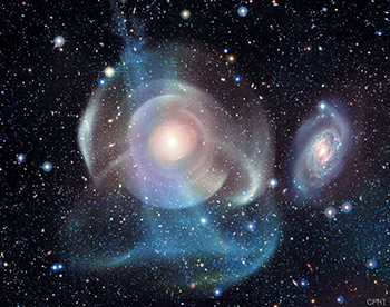
IMAGE CREDIT: CFHT, COELUM, MEGACAM, J.-C. CUILLANDRE (CFHT) & G. A. ANSELMI (COELUM)
The Webb Telescope will study galaxies as they are forming, understand the formation of stars and planets, and directly image novas and planets outside our solar system. Shown, an image of galaxy NGC 474, captured by the Canada-France-Hawaii Telescope (CFHT), which is on the summit of Mauna Kea in Hawaii. [CREDIT] CFHT et. al.;
“We have two spectacular presenters to take us through the brave new world of imaging from the cosmos to the molecule,” said NIH Director Francis Collins, who was on hand to introduce the speakers. “You are about to travel through 36 orders of magnitude. Put on your seat belts, and let’s see where it takes us.”
Imaging the Cosmos: Mather, a senior astrophysicist in the Observational Cosmology Laboratory at NASA’s Goddard Space Flight Center (Greenbelt, Maryland), shared the 2006 Nobel Prize in physics (with George Smoot) for the “discovery of the blackbody form and anisotropy of the cosmic microwave background” using the Cosmic Background Explorer (COBE) satellite launched by NASA in 1989. This discovery is in essence the afterglow of the Big Bang 13.8 billion years ago that gave birth to the universe. Mather was a key figure leading the COBE space mission and the principal investigator for the far-infrared absolute spectrophotometer on COBE.
“The sky is filled with at least 100 billion galaxies,” said Mather. Each galaxy “is 100 billion stars orbiting a common center pulled together by gravity.” And the universe is expanding. Astronomers can measure this expansion based on the wavelength of the light emitted from distant stars.
The Hubble Space Telescope, which was launched into low Earth orbit (at an altitude of 340 miles) in 1990, was intended to help astronomers “see the first galaxies forming,” said Mather. But it wasn’t strong enough. So now NASA, in collaboration with the European Space Agency and the Canadian Space Agency is building the James Webb Space Telescope. (Mather is the senior project scientist.) This large infrared telescope has a mirror that is in 18 hexagonal segments and measures 21 feet in diameter, about 2.5 times as long as Hubble’s mirror, folds up to fit in a rocket, and will take two weeks to unfold. It is scheduled to launch in 2020 and will be about 930,000 miles beyond Earth. Astronomers are excited that the Webb telescope will be used to study galaxies as they are forming, understand the formation of stars and planets, and directly image novas and planets outside our solar system.
Imaging Within: While Mather is looking skyward, Jennifer Lippincott-Schwartz is looking inward—at the tiniest elements of cells. Before becoming a senior group leader at the Howard Hughes Medical Institute (HHMI) Janelia Research Campus (Ashburn, Virginia) in 2016, she was a senior investigator in the National Institute of Child Health and Human Development (NICHD). She and George Patterson, then a postdoc in her lab and now a senior investigator in the National Institute of Biomedical Imaging and Bioengineering, created a photoactivatable form of green fluorescent protein that can be switched on using flashes of light. She hosted Eric Betzig (now at HHMI) and his colleague in her NICHD lab so they could build a super-resolution microscope called photoactivated localization microscopy (PALM), for which Betzig won the 2014 Nobel Prize in chemistry.

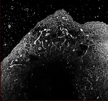
IMAGE COURTESY OF JENNIFER LIPPINCOTT-SHWARTZ, WES LEGANT, AND ERIC BETZIG, HHMI
Super-resolution microscopy allows biologists to see deep inside of cells and produce images that rival those of the universe. Shown: The Lattice Light-Sheet Microscope can do single-molecule imaging of lipids in membranes.
Lippincott-Schwartz explained that light microscopes have resolution limits—based on the wavelength of light, refractive index of the medium, and angle of the converging spot—and can’t image many cell details. Super-resolution microscopy, such as PALM and the lattice light-sheet microscope (also developed by Betzig), however, can image the molecular complexity of the subcellular landscape. In PALM, a fluorophore is used as a point source of light, which Lippincott-Schwartz compared to stars. She calls it “super-resolution by pointillism.” She has begun using even more advanced super-resolution microscopy techniques to explore the inner workings of cells.
Imaging Life and the Cosmos: Both scientists acknowledge the commonalities their two fields share. Mather said he’s grateful for the work of biologists who push the boundaries of microscopy. Lippincott-Schwartz pointed out that high-resolution microscopy has benefited from innovations in telescopes such as deformable mirrors that compensate for wave-front distortions in light coming from stars.
“We share the common intellectual tools trying to understand instrumentation [and] the objects we’re looking at,” said Lippincott-Schwartz. Whether it’s looking at fluorophores in PALM or stars in the solar system, “[we] use the same algorithms for trying to understand the distribution and behavior of these systems.”
You can watch the March 13, 2018, videocast online at https://videocast.nih.gov/launch.asp?23753. To find out more about the DeMystifying Medicine lecture series, go to https://demystifyingmedicine.od.nih.gov/DM18/TopicIntros18.html.
This page was last updated on Thursday, April 7, 2022
