NIH Microscopy Lights Up Dulles Airport
Spectacular Mega-magnified Images Reveal Cellular Mysteries
Brains, bone, muscle, blood—along with fish fins and flower parts—are among the biological players whose portraits light up the C-concourse walkway at Washington Dulles International Airport. These images—most of which are from scientists at or supported by NIH—comprise Life: Magnified, a gallery exhibit that showcases backlit, mega-magnified images of cells and other microscopic biological structures.
The show runs through November and is co-sponsored by the National Institute of General Medical Sciences, the American Society for Cell Biology, and the Metropolitan Washington Airports Authority’s Arts Program. More than a million ticketed travelers will see the exhibit at Dulles. It’s likely that even more people will see it on a screen. The online version of Life: Magnified is at http://www.nigms.nih.gov/education/life-magnified/, where users can read enhanced captions and freely download high-resolution versions of all of the images for research, education, and other noncommercial purposes.
In addition, news of the exhibit has been picked up by various scientific journals and media outlets including the Washington Post, Scientific American, National Geographic, The Atlantic, NBC News Online, and even BuzzFeed, and My Modern Met.
Of the 46 images in the collection (selected from more than 600 submissions), 11 are from intramural labs: National Institute of Allergy and Infectious Diseases (NIAID); Eunice Kennedy Shriver National Institute of Child Health and Human Development (NICHD); National Heart, Lung, and Blood Institute (NHLBI); and the National Eye Institute (NEI). About 20 different organisms are represented in the exhibit, including humans, common biomedical models, a few more exotic creatures—a gecko, an anglerfish, and a lone star tick—and eight pathogens.
The exhibit organizers hope that after the exhibit is over, some of the images will be displayed on the NIH campus.
The following images were produced by the NIH intramural program.
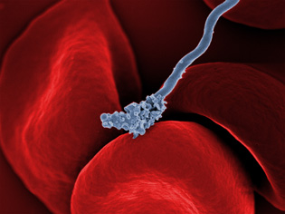
TOM SCHWAN, ROBERT FISCHER, AND ANITA MORA, NIAID
Relapsing fever bacterium (gray) on red blood cells: The long, spiral-shaped bacterium (gray) in this image causes relapsing fever, a disease characterized by recurring high fevers, muscle aches, and nausea. The relapses result from the bacterium's unusual ability to change the molecules on its outer surface, allowing it to dodge the human immune system. The disease is transmitted through the bite of a tick (not the same species that transmits Lyme disease) and is found in parts of the Americas, the Mediterranean, central Asia, and Africa.
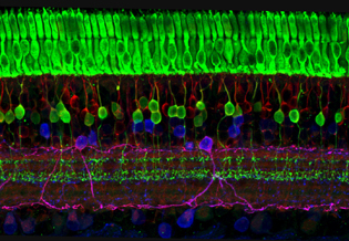
WEI LI, NEI
The eye uses many layers of nerve cells to convert light into sight: This image captures the many layers of nerve cells in the retina. The top layer (green) is made up of cells called photoreceptors that convert light into electrical signals to relay to the brain. The two best-known types of photoreceptor cells are rod- and cone-shaped. Rods help us see under low-light conditions but can’t help us distinguish colors. Cones don’t function well in the dark but allow us to see vibrant colors in daylight.
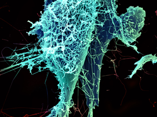
HEINZ FELDMANN, PETER JAHRLING, ELIZABETH FISCHER, AND ANITA MORA, NIAID
Stringlike Ebola virus peeling off an infected cell:
After multiplying inside a host cell, the stringlike Ebola virus is emerging to infect more cells. Ebola is a rare, often fatal disease that occurs primarily in tropical regions of sub-Saharan Africa. The virus is believed to spread to humans through contact with wild animals, especially fruit bats. It can be transmitted between one person and another through bodily fluids.
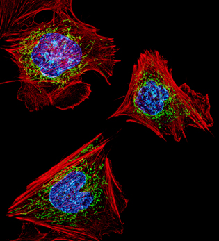
DYLAN BURNETTE AND J. LIPPINCOTT-SCHWARTZ, NICHD
Cells with nuclei in blue, energy factories in green, and the actin cytoskeleton in red: The cells shown here are fibroblasts, one of the most common cells in mammalian connective tissue. These particular cells were taken from a mouse. Scientists used them to test the power of a new microscopy technique that offers vivid views of the inside of a cell. The DNA within the nucleus (blue), mitochondria (green), and cellular skeleton (red) are clearly visible.
PAKORN KANCHANAWONG, NATIONAL UNIVERSITY OF SINGAPORE AND NHLBI; CLARE WATERMAN, NHLBI
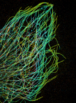
Tiny strands of tubulin, a protein in a cell’s skeleton: Just as our bodies rely on bones for structural support, our cells rely on a cellular skeleton. In addition to helping cells keep their shape, this cytoskeleton transports material within cells and coordinates cell division. One component of the cytoskeleton is a protein called tubulin, shown here as thin strands.
DANIELA MALIDE, NHLBI
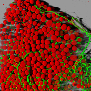
Fat cells (red) and blood vessels (green): A mouse's fat cells (red) are shown surrounded by a network of blood vessels (green). Fat cells store and release energy, protect organs and nerve tissues, insulate us from the cold, and help us absorb important vitamins.
ROBERT HEINZEN, ELIZABETH FISCHER, AND ANITA MORA, NIAID
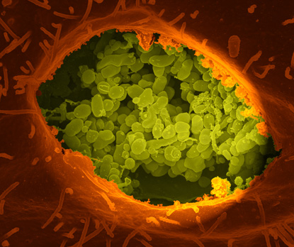
Q-fever bacteria (yellow) in an infected cell: This image shows Q-fever bacteria (yellow), which infect cows, sheep, and goats around the world and can infect humans as well. When caught early, Q fever can be cured with antibiotics. A small fraction of people develop a more serious, chronic form of the disease.
NASSER RUSAN, NHLBI
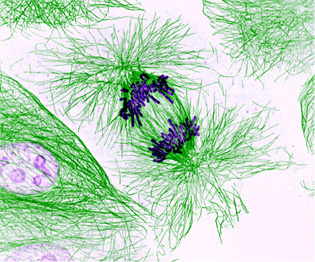
Dividing cells showing chromosomes (purple) and cell skeleton (green): This pig cell is in the process of dividing. The chromosomes (purple) have already replicated, and the duplicates are being pulled apart by fibers of the cell skeleton known as microtubules (green). Studies of cell division yield knowledge that is critical to advancing understanding of many human diseases, including cancer and birth defects.
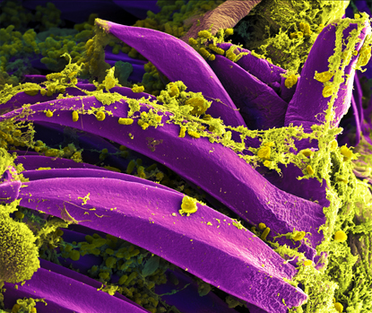
B. JOSEPH HINNEBUSCH, ELIZABETH FISCHER, AND AUSTIN ATHMAN, NIAID
Bubonic plague bacteria (yellow) on part of the digestive system in a rat flea (purple): Here, bubonic plague bacteria (yellow) are shown in the digestive system of a rat flea (purple). Carried by rodents and spread by fleas, the bubonic plague killed a third of Europeans in the mid-14th century. Today, it is still active in Africa, Asia, and the Americas, with as many as 2,000 people infected worldwide each year. If caught early, bubonic plague can be treated with antibiotics.
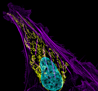
DYLAN BURNETTE AND J. LIPPINCOTT-SCHWARTZ, NICHD
Bone cancer cell (nucleus in light blue): This image shows an osteosarcoma cell with DNA in blue, energy factories (mitochondria) in yellow, and actin filaments, part of the cellular skeleton, in purple. One of the few cancers that originate in the bones, osteosarcoma is extremely rare, with fewer than a thousand new cases diagnosed each year in the United States.
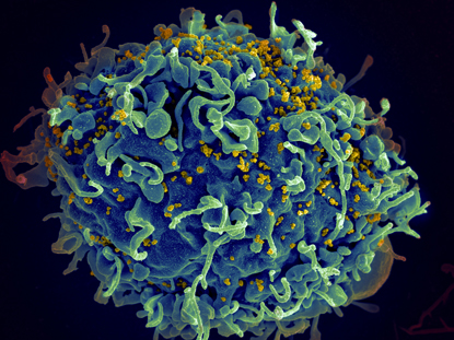
SETH PINCUS, ELIZABETH FISCHER, AND AUSTIN ATHMAN, NIAID
HIV, the AIDS virus (yellow), infecting a human cell: This human T cell (blue) is under attack by the human immunodeficiency virus (yellow), the virus that causes AIDS. The virus specifically targets T cells, which play a critical role in the body’s immune response against invaders like bacteria and viruses.
This page was last updated on Wednesday, April 27, 2022
