NIH in History
History Mystery Solved
Discovery that Revolutionized Epithelial Cell Research
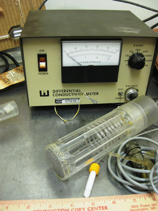
No, it wasn’t a prototype for a flux capacitor.
The “History Mystery” photo that appeared in the May-June issue of the NIH Catalyst (https://irp.nih.gov/catalyst/v21i3/nih-in-history) elicited 14 responses to our plea for help in identifying the equipment used by the late Roderic E. Steele, a researcher in the National Heart, Lung, and Blood Institute (NHLBI) from 1975 to 1988. Three were whimsical guesses—a flux capacitor (part of the time-travel machine featured in the Back to the Future trilogy); an early breast pump; and a device to deliver electroshock therapy. But most respondents provided real clues. They gave us contacts, descriptions, and journal articles. We thank everyone who helped identify this object. Now we know it is a “keeper” for the NIH Stetten Museum collection.

MICHELE LYONS, OFFICE OF NIH HISTORY
NIH scientists helped the NIH Office of History discover that this conductivity meter was part of a system that the late NHLBI scientist Roderic E. Steele started developing in the 1970s: a porous-bottom culture dish that revolutionized research on epithelial cells.
So what is it? It turns out that it’s equipment for a porous-bottom culture dish (PBCD) that Steele helped to develop for research on epithelial cells.
“This product truly revolutionized the way the epithelial cell community studied cells, particularly for performing transport assays,” said Gregory Germino, Deputy Director of the National Institute of Diabetes and Digestive and Kidney Diseases (NIDDK). Transport assays measure the movement of materials across epithelial layers. “The conductivity meter would have been used to measure resistance, a measure of how ‘tight’ the cell junctions had become, and also how much ionic activity could be induced by various interventions.”
Epithelial cells form one or more layers on internal and external surfaces of the body. They line the major cavities, most organs, and blood vessels inside the body and the skin on the outside. They are tightly packed together and have two membranous surfaces: the apical (upper) and the basal (lower). Molecules—including nutrients, hormones, growth factors, ions, and oxygen as well as carbon dioxide and other waste products—are transported through the membranes. But such transport is difficult to measure when the cells are grown in a normal culture dish: There’s a lack of access to the basal surfaces of cells which are against the solid plastic of the tissue culture dish. The porous bottom of Steele’s culture dish, however, simulated a more natural environment. It allowed for the passage of transported solutes and provided a conductive pathway for electrical measurements.
Steele’s journey to create the PBCD began with a paper that he and colleagues published in 1977 on work he did at Stanford University (Stanford, Calif.). He was studying sodium transport and carbon dioxide production to determine how the chemical energy of cellular metabolism is converted to the electrochemical energy of active transport. Instead of using a conventional culture dish, he fashioned a porous-bottom dish out of a toad bladder. The bladder was cut in half and suspended, like a tiny trampoline, across a special glass chamber designed to simultaneously measure sodium transport and oxygen consumption (J Membr Biol 34:289–312, 1977).
By the spring of 1983, Steele was working with T. Andrew Guhl, an inventor and supervisor at Becton Dickinson, on commercializing the PBCD. In February 1984, Becton Dickinson was ready to take their commercial version of Steele’s handmade setup to outside researchers for evaluation. Guhl wrote in a letter to Steele, “I envision this particular product to be the first of a family of products incorporating a porous membrane for cell culture experiments involving feeding of the cell monolayer from the basal-lateral surface.”
In 1986, Steele published a paper with Joseph S. Handler (NHLBI) in the American Journal of Physiology in which they described four porous bottoms they had tried—cellulose ester, polycarbonate, collagen, and placental amnion. The first three types were attached to a polycarbonate ring that formed the sides. The amnion bottoms were tucked between two hollow polyethylene stoppers stacked on top of each other in an “embroidery-hoop arrangement” (Am J Physiol 251:C136–C139, 1986, and Methods Enzymol 171:736–744, 1989).
The group continued their development of the PBCD, publishing an article in 1992 about a more complicated set-up (J Tissue Cult Methods 14:259–264, 1992).
After Steele left NIH in 1988, he continued his association with Handler’s lab. He eventually retired to California, where he died in 2011. His widow told me that she did not really understand what he was working on and didn’t have any papers or objects to document his work. We were fortunate, however, that a few letters and handwritten data sheets mailed between Steele and Guhl were tucked into the box containing the PBCD. Usually, we are not so lucky as to have any of the correspondence between the federal employee inventor and the commercial operation.
The expertise of the scientific community is an important resource for the NIH Stetten Museum. The museum’s collection covers all NIH history and research, so staff members are sometimes unfamiliar with equipment that scientists and technicians recognize immediately. Without your help, we would never be able to identify everything in our collection. It’s a good thing we like to learn.
HERE ARE SOME OF THE RESPONSES WE RECEIVED:
Victoria Hampshire, FDA
I worked in Dr. Steele’s laboratory from May 1982 to about May 1983. I was a technologist. I moved on to veterinary school and lost touch but returned to the area to work and by that time he had moved on back to California. I don’t recognize the pictures in the Catalyst but I knew that Dr. Steele was interested in talking to Becton Dickinson.
Sarah Sohraby, NEI
Bob Balaban (NHLBI), Rick Fisher (NEI), Mark Knepper (NHLBI), and I were all working in NHLBI’s Laboratory of Kidney and Electrolyte Metabolism, along with Rod Steele. The porous membrane shown may well be a system that Bob was trying to develop to do mass cultures of cells that would remain well perfused and oxygenated to be used in nuclear magnetic resonance experiments. These systems would get easily contaminated.
Michael Gottesman, Deputy Director for Intramural Research
I would guess that the conductivity meter is used to measure resistance across cell monolayers to determine whether they are forming tight junctions to allow studies on trans-epithelial transport. The equipment looks jury-rigged; modern versions of this are smaller and less interesting-looking.
Maurice Berg, NHLBI
The project was measuring the trans-epithelial transport through a layer of tissue culture cells on a porous support. The conductivity meter in the photo was used to measure the transport characteristics. I do not see a holder for the porous support.
Jay Knutson, NHLBI
I arrived early in 1984, and I recall Rod giving lab-meeting lectures about his PBCDs, but I can’t say with certainty the meter shown was used for those.
Gregory Gemino, NIDDK
I believe Joe Handler may have been developing transwell chambers for growth of polarized epithelial cells back then. This product truly revolutionized the way the epithelial cell community studied cells, particularly for performing transport assays. The conductivity meter would have been used to measure resistance, a measure of how “tight” the cell junctions had become, and also how much ionic activity could be induced by various interventions.
Edward Kerns, NCATS
Not long after Dr. Steele’s article, other articles started to appear from the lab of Ron Borchardt (University of Kansas in Lawrence) describing intestinal transport experiments with a porous polycarbonate membrane, (similar to that described by Dr. Steele). Whether Drs. Steele and Borchardt knew of each other’s research is not known to me. The cell line used most often for this type of experiment is Caco-2 and it is a key assay used today for drug permeability. During such studies, it is common to test for confluence of the monolayer of cells on the permeable membrane and this is done with a conductivity meter, an early version of which is shown in the picture. Another clue is that Becton Dickinson is a current manufacturer of porous membrane cell culture plates for cell layer drug permeability studies. I was not, however, familiar with Dr. Steele or his work at NIH.
Mark Knepper, NHLBI
Epithelial biologists have long recognized that epithelial cells grown in culture do not polarize and differentiate well when grown on solid surfaces. Apparently, essential nutrients and growth factors cannot reach the basal aspect of cells in culture as easily as when cells are in their natural setting. In the early 1980s, this problem stimulated Dr. Joseph Handler, a pioneer in epithelial physiology and a member of NHLBI’s Laboratory of Kidney and Electrolyte Metabolism (LKEM), to seek out Dr. Roderic Steele of NHLBI’s Laboratory of Technical Development (LTD) to seek a solution. Steele invented a device with a porous bottom surface on which to grow the epithelial cells. This device was found to foster the normal process of epithelial differentiation, opening the door for rapid progress in epithelial physiology. Today, investigators worldwide routinely grow epithelial cells on commercial “descendants” of Dr. Steele’s invention. Incidentally, this is one of many collaborations between LTD (led by Dr. Robert Bowman) and LKEM (led by over the years by Robert Berliner, Jack Orloff, Maurice Burg, and myself). LTD and LKEM were among the first labs created when NHLBI was established (as the National Heart Institute) on the Bethesda campus in the late 1940s.
This page was last updated on Thursday, April 28, 2022
