Colleagues: Recently Tenured
Carson Chow, Ph.D., NIDDK
Senior Investigator, Laboratory of Biological Modeling

Education: University of Toronto, Toronto (B.A.S. in engineering science); Massachusetts Institute of Technology, Cambridge, Mass. (Ph.D. in physics)
Training: Department of Astrophysical, Planetary and Atmospheric Sciences at the University of Colorado, Boulder; Neuromuscular Research Center and the Department of Mathematics at Boston University
Before coming to NIH: Associate professor of mathematics and of neurobiology at the University of Pittsburgh
Came to NIH: In 2004
Selected professional activities: Action editor for Journal of Computational Neuroscience; associate editor for Journal of Applied Mathematics
Outside interests: Skiing, cycling, playing golf and tennis; spending time with daughter; blogging at http://sciencehouse.wordpress.com
Research Interests: I am interested in mathematical and computational biology as applied to neuroscience, metabolism, obesity, gene regulation, and population genetics. I construct reduced models of complex systems that can be analyzed mathematically to answer specific questions. The interdisciplinary nature of my research involves collaborations with other labs at NIH. For example, with Kevin Hall (NIDDK), I used a simple model of human body weight change to show that the increase in the U.S. food supply over the past 30 years explains the obesity epidemic. With Stoney Simons (NIDDK), I used simple concepts from the mathematical theory of groups to make testable predictions for molecular mechanisms involved in steroid-mediated gene induction. In complex neural disorders such as autism, it is difficult to understand the connection between genotype and phenotype because genes act at the molecular level while disorders are manifested at the behavioral level. I will use computational models to analyze how molecular perturbations affect the operation of neural circuits, associate their behavior with symptoms, and collaborate with NIMH labs to obtain imaging data.
Serena M. Dudek, Ph.D., NIEHS
Senior Investigator, Synaptic and Developmental Plasticity Group, Laboratory of Neurobiology
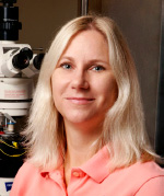
Education: University of California at Irvine (B.S. in biology); Brown University, Providence, R.I. (Ph.D. in neuroscience)
Training: Postdoctoral training in the Department of Neurobiology at the University of Alabama at Birmingham; Laboratory of Cellular and Synaptic Neurophysiology, NICHD
Came to NIH: In 1996 for training; appointed tenure-track investigator in 2001
Other professional activities: Edited book titled Transcriptional Regulation by Neuronal Activity; member of The Society for Neuroscience Program Committee
Outside interests: Volunteering as a paramedic; observing the neural development of her two-year-old daughter
Research Interests: Our group studies the regulation of synaptic effectiveness and how synaptic changes early in development are consolidated to last a lifetime. During postnatal development, mammals, including humans, acquire vast amounts of information by interacting with their environments. In contrast to creatures with nervous systems that are fully prewired at birth, mammals benefit from an enormous flexibility in behavior because of the driving force of experience on their brain development. This flexibility comes at a potential cost, however, because interactions with noxious or abnormal environments can cause lasting and often deleterious changes in brain circuitry.
Using patch clamp and extracellular recordings in brain slices, confocal microscopic imaging, and molecular and cellular techniques, our group determines how the connections (synapses) in the brain change or are pruned in response to neuronal activity. Such pruning regulates the critical periods of postnatal development when plasticity (the ability of the brain to reorganize itself in response to the environment, experiences, and outside influences) is most robust, and our research gives clues as to why some brain regions are more plastic than others. Recently, our exploration of plasticity modulators has led us to some interesting discoveries in a previously unappreciated region of the brain, hippocampal area CA2, which we speculate could be important in several psychiatric disorders. By having a better understanding of how environmental factors play a role in forming the circuitry of the brain, we hope to address the associated problems of brain disease caused by toxicant exposure.
Patrick E. Duffy, M.D., NIAID
Senior Investigator; Chief, Laboratory of Malaria Immunology and Vaccinology
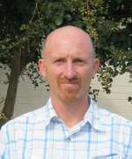
Education: United States Military Academy, West Point, N.Y. (B.S. in civil engineering with concentrations in English literature and basic sciences); Duke University School of Medicine, Durham, N.C. (M.D.)
Training: Residency in internal medicine at Walter Reed Army Medical Center (Washington, D.C.); medical research fellowship at Walter Reed Army Institute of Research; postdoctoral training in molecular parasitology malaria research at NIAID’s Laboratory of Malaria Research
Before coming to NIH: Director of the malaria program at the Seattle Biomedical Research Institute and affiliate professor of global health at the University of Washington (Seattle); director of preclinical vaccine development for the malaria program at the Walter Reed Army Institute of Research
Came to NIH: In 1991 for training; in November 2009 returned as chief of the Laboratory of Malaria Immunology and Vaccinology
Selected professional activities: Long-standing organizer of the East African Regional Training Workshop series for young scientists working on protozoan pathogens; senior investigator for large longitudinal cohort studies in Tanzania and Mali
Outside interests: Jogging; traveling
Research Interests: We conduct basic, translational, and clinical research to develop malaria vaccines. Malaria is caused by a parasite that is transmitted by mosquitoes to humans. We study malaria pathogenesis and immunity with a focus on pregnant women and infants. We performed seminal studies in Kenya that identified a distinct parasite phenotype that causes malaria in pregnant women. We are also investigating the pathogenesis of severe malaria in children.
Our central product development unit, which operates more like a small biotech firm than a typical research laboratory, makes prototype malaria vaccines. We also formulate antigens; develop assays and animal trials that define the potential for protection; and establish clinical trials to test vaccines in the United States and in the developing world.
We are developing two kinds of malaria vaccines: a pregnancy malaria vaccine (PMV) and a vaccine that interrupts malaria transmission (VIMT). The PMV blocks the surface proteins on infected red cells that the parasite uses to bind inside the placenta. The VIMT elicits immune responses that destroy malaria parasites as they enter either the mosquito or the human host. Our lab’s overarching goal is to protect children and pregnant women from malaria and to eliminate the disease from low-transmission areas of the world.
Daniel Fowler, M.D., NCI-CCR
Senior Investigator; Head, Cytokine Biology Section, Experimental Transplantation and Immunology Branch

Education: Kalamazoo College, Kalamazoo, Mich. (B.A. in health sciences); Wayne State University School of Medicine, Detroit (M.D.)
Training: Residency in internal medicine and pediatrics at Detroit Medical Center (Detroit); training in medical oncology at NCI
Came to NIH: In 1990 for training; became investigator in 1994; appointed tenure-track investigator in 1999
Selected professional activities: Member of the American Society of Blood and Marrow Transplantation and the American Society of Clinical Investigation
Outside interests: Parenting; playing sports; landscaping
Research Interests: My research focuses on T-cell and T helper (Th) cell regulation after allogeneic (from different but matched donors) transplantation of stem cells. In animal models, we found that donor Th1 cells mediated graft-versus-host disease (GVHD), the main transplant complication, whereas Th2 cells inhibited GVHD. In 1999, in collaboration with the Department of Transfusion Medicine, I developed a method to produce human Th2 cells, filed an Investigational New Drug application with the FDA, and implemented a trial of Th2 cell therapy at the Clinical Center in NCI’s Experimental Transplantation and Immunology Branch.
Recently, a student gave me a coffee mug depicting the mTOR (mammalian target of rapamycin) signaling pathway to symbolize my fixation on the immune suppression drug rapamycin. We found that Th2 cells develop resistance to rapamycin. In animal models, rapamycin-resistant Th2 cells prevented GVHD and graft rejection more effectively than did control Th2 cells.
In 2004, I initiated a second-generation clinical trial using rapamycin-resistant Th2 cells. Ongoing results indicate that such T-rapa cells safely prevent graft rejection in the “mini-transplant” setting (using low-intensity, outpatient-dose chemotherapy) and mediate graft-versus-tumor effects with reduced GVHD. In our current research, we seek to further define the role of Th1 and Th2 cell manipulation in transplantation therapy for cancer.
Kevin D. Hall, Ph.D., NIDDK
Senior Investigator, Laboratory of Biological Modeling
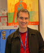
Education: McMaster University, Hamilton, Ont. (B.S. in physics); McGill University, Montreal (Ph.D. in physics)
Before coming to NIH: Scientist at Entelos Inc. (Menlo Park, Calif.)
Came to NIH: In 2003
Outside interests: Cycling; hiking; scuba diving; motorcycling; playing guitar
Research Interests: My laboratory studies mammalian metabolism, body-weight regulation, and the physiological dysregulation that occurs in obesity, diabetes, anorexia, and cachexia (physical wasting and malnutrition associated with chronic disease). We study humans and rodents to better understand the complex mechanisms regulating macronutrient metabolism, body composition, and energy expenditure. We use mathematical models to quantitatively describe, explain, integrate, and predict our experimental results. We have created several models ranging from a single equation that describes the proportion of weight changes attributable to body fat to computational models representing the dynamics of whole body metabolism, including body composition change and metabolic fluxes. We are also developing new methods for measuring food-intake behavior over extended time periods as well as practical tools to help predict weight changes resulting from obesity interventions at the individual and population levels.
David B. Sacks, M.B.Ch.B., F.R.C.Path., CC
Senior Investigator; Chief, Clinical Chemistry
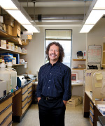
Education: University of Cape Town, Cape Town, South Africa (M.B., Ch.B.)
Training: Residency in internal medicine at Georgetown University–affiliated hospitals (Washington, D.C.); residency in clinical pathology and fellowship training in clinical pathology and clinical chemistry at Washington University School of Medicine (St. Louis)
Before coming to NIH: Associate professor of pathology at Harvard Medical School (Boston); Medical Director of Clinical Chemistry and Director of the Clinical Pathology Training Program at Harvard’s Brigham and Women’s Hospital (Boston)
Came to NIH: In January 2011 as Chief of Clinical Chemistry
Outside interests: Listening to music; jogging; watching rugby
Research Interests: We are investigating the derangements of calcium and calmodulin signaling in disease. Calmodulin is a ubiquitous, highly conserved protein that plays a critical role in many essential cellular functions. A considerable body of evidence implicates calcium and calmodulin in tumorigenesis. Several calmodulin targets, such as the estrogen receptor (ER) and IQGAP1, contribute to tumorigenesis. We have shown that calmodulin binds to ER. This interaction both enhances the stability of ER and is required for ER-mediated transcriptional activity in the nucleus.
Calmodulin also regulates IQGAP1 function. IQGAP1 is a scaffolding protein that assembles multiprotein complexes and integrates signaling cascades. We showed that IQGAP1 binds the human epidermal growth factor receptor (HER2) and several components of the mitogen-activated protein kinase pathway. We identified several important functions of IQGAP1: It promotes cell motility, neurite outgrowth, and cell adhesion, and it participates in microbial pathogenesis. We documented that IQGAP1 enhances breast tumorigenesis. Overexpression of IQGAP1 is observed in several tumors; both calmodulin and IQGAP1 concentrations are increased in highly metastatic cells. We are elucidating the molecular mechanisms by which IQGAP1 integrates signaling pathways and promotes malignant transformation.
Mark Stopfer, Ph.D., NICHD
Senior Investigator and Head, Unit on Sensory Coding and Neural Ensembles
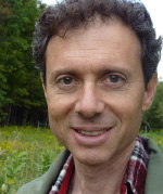
Education: Yale University, New Haven, Conn. (B.S. in biology; M.S., M.Phil., and Ph.D. in psychology)
Training: California Institute of Technology (Pasadena, Calif.)
Before coming to NIH: Postdoctoral training at the California Institute of Technology
Came to NIH: In 2002
Outside interests: Playing guitar; feeding birds
Research Interests: I’m interested in the brain mechanisms that gather and organize sensory information to build transient and sometimes enduring internal representations of the environment. Information does not flow passively from the outer environment through neural circuits, coming to rest as memories or actions. Rather, neural circuits process and dramatically transform information in myriad ways, providing multiple advantages to the animal. Using relatively simple animals, including Drosophila, our lab combines electrophysiological, anatomical, genetic, behavioral, computational, and other techniques to examine the ways that sensory stimuli drive neural circuits to process information. Recently, we’ve investigated several related questions: What mechanisms, including the transient oscillatory synchronization and slow temporal firing patterns of ensembles of neurons, underlie information coding and decoding? How are natural features of sensory stimuli extracted? How do sensory systems develop, how are innate sensory preferences determined, and how does innate information within the nervous system differ from learned information?
Terry A. Van Dyke, PH.D., NCI-CCR
Senior Investigator; Head, Cancer Pathways and Mechanisms; Director, Mouse Cancer Genetics Program; Director, Center for Advanced Preclinical Research
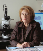
Education: University of Florida, Gainesville, Fla. (B.S. in interdisciplinary studies; Ph.D. in medical sciences)
Training: Postdoctoral training at the University of Florida, Stony Brook University (Stony Brook, N.Y.), and Princeton University (Princeton, N.J.)
Before coming to NIH: Professor, Department of Genetics and Biochemistry and Biophysics, University of North Carolina at Chapel Hill (Chapel Hill, N.C.)
Came to NIH: In September 2007
Outside interests: Walking in the woods and other natural areas; playing the clarinet; listening to music
Research Interests: Our lab studies the mechanisms and pathways to cancer development at the genetic, molecular, cellular, and organ levels. Because cancer can develop in more than 100 mammalian cell types, and does so amidst complex cell-cell and cell-environment interactions, we have used genetically engineered mice (GEM) as the foundation for our analyses. We have established several preclinical cancer models that have facilitated analyses of the tumor suppressors p53, pRb, and PTEN, among others. Using GEM, we can do studies that are not possible in humans such as a detailed examination of the molecular and cellular events in developing tumors. We couple in vivo approaches with in vitro primary cell culture approaches to refine our discoveries.
Our projects have clarified aspects of cancer cell proliferation, apoptosis, and invasion. Studies are under way to define the chromosomal and gene-expression aberrations that characterize these events. We are also exploring the mechanisms of angiogenesis and invasiveness. We have developed preclinical models for cancers of the breast, prostate, ovary, and brain. These models are also being used to develop live-animal-imaging methodologies to characterize the disease process and monitor preclinical therapeutic testing. Thus, our lab uses a toolbox of modern technologies to approach the complexities of this aggressive and devastating disease.
Di Xia, Ph.D., NCI-CCR
Senior Investigator; Head, Crystallography Section, Laboratory of Cell Biology

Education: Zhejiang University, Hangzhou, China (B.S. in chemistry); Purdue University, West Lafayette, Ind. (Ph.D. in structural biology)
Training: Postdoctoral training in membrane protein structural biology at the University of Texas, Southwestern Medical Center at Dallas (Dallas)
Before coming to NIH: Assistant lecturer, Department of Biochemistry, University of Texas, Southwestern Medical Center at Dallas
Came to NIH: In 1998
Outside interests: Spending time with wife and son; reading
Research Interests: Our lab studies membrane proteins, their atomic structure, and their role in multidrug resistance. Membrane proteins are important for cell-cell communication, recognition, adhesion, and membrane fusion; material exchange, transportation, and detoxification; and cellular energy conservation.
In this era of structural genomics, we have determined the three-dimensional structure of many proteins. But in spite of intense efforts, scientists have so far been able to decipher the structure of only a few membrane proteins. We use molecular, biological, and crystallographic methods to obtain atomic resolution structures of a few selected families of membrane proteins in order to understand how they function. But it’s often difficult to purify large enough quantities of these proteins to yield an adequate number of crystals for analysis.
We are particularly interested in various membrane transporters that play important roles in drug resistance and in pumping protons. We are studying membrane transport proteins called ABC transporters (for ATP-binding cassette transporters). Our studies have helped to elucidate mechanisms of functions at near-atomic resolution of these membrane transporters and provided structural information essential for understanding the interactions of these proteins with various drugs.
Terry P. Yamaguchi, Ph.D., NCI-CCR
Senior Investigator; Head, Cell Signaling and Vertebrate Development Section
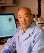
Education: University of Toronto, Toronto (B.Sc. in pharmacology and physiology; M.Sc. in anatomy and cell biology; Ph.D. in molecular and medical genetics)
Training: Postdoctoral fellow at Harvard University (Cambridge, Mass.)
Came to NIH: In 2000
Outside interests: Organizing, playing, and coaching ice hockey; playing volleyball; running; playing keyboards and guitar; entertaining son and daughter
Research Interests: Our lab studies Wnt proteins, which are powerful signaling molecules that control the growth, differentiation, and movement of embryonic and adult stem cells. Genetic mutations in Wnt signaling pathways can lead to unrestrained signaling and cancer. We are particularly interested in the molecular mechanisms of Wnt signaling during development and its implications for diseases such as cancer. We have uncovered the genetic regulatory networks that control the formation of multipotent mesoderm progenitors during gastrulation, an early phase in embryonic development. The mesoderm—the middle layer of an embryo’s three germ layers that form during gastrulation—undergoes an epithelial-to-mesenchymal transition (EMT) as it differentiates into connective tissue, bone and cartilage, muscle, blood and blood vessels, and other organs. We have identified Wnt-dependent transcription factors that control EMT as well as the specification and maturation of mesodermal progenitors. Because Wnt signals and EMT play critical roles in tumorigenesis and metastasis, future studies will address the role that embryonic Wnt target genes play in adult tumors caused by disregulated Wnt signaling.
Victor Zhurkin, Ph.D., NCI-CCR
Senior Investigator; Head, DNA and Nucleoproteins Section, Laboratory of Cell Biology
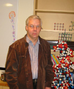
Education: Lomonosov Moscow State University, Moscow (M.S. in applied mathematics); Moscow Physical-Technical Institute, Moscow (Ph.D. in molecular biophysics); Lomonosov Moscow State University (D.Sc., in molecular biophysics)
Training: Postdoctoral training at Engelhardt Institute of Molecular Biology (Moscow)
Came to NIH: In 1989
Research Interests: We are interested in the structural aspects of protein-DNA interactions in chromatin, where DNA is accessible to the transcription factors. One such factor is the p53 protein, which mediates the transcription of hundreds of human genes and plays an important role as a tumor suppressor in human cancers.
We found that p53 can bind to its response element when it is properly exposed in the nucleosome (DNA sequence wrapped around a core of proteins called histones). The physiological importance of this result is best illustrated by comparing p53’s high affinity to the response elements associated with cell-cycle arrest (CCA sites) with its low affinity to the sites associated with apoptosis (Apo sites). We predicted and found that the CCA sites are likely to be exposed on the nucleosome surface. In contrast, the Apo sites are often hidden inside. The difference may be key to p53-DNA binding and the kinetics of transactivation of the corresponding genes.
In the near future, we will consider other genes that are characterized by various kinetics of p53-induced activation. The results of bioinformatic analysis should give us a better understanding of the relationships between the mechanisms of gene regulation and the genomic environment of the p53 sites.
If you have been tenured in the last year or so, The NIH Catalyst will be in touch soon to include you on these pages.
This page was last updated on Monday, May 2, 2022
