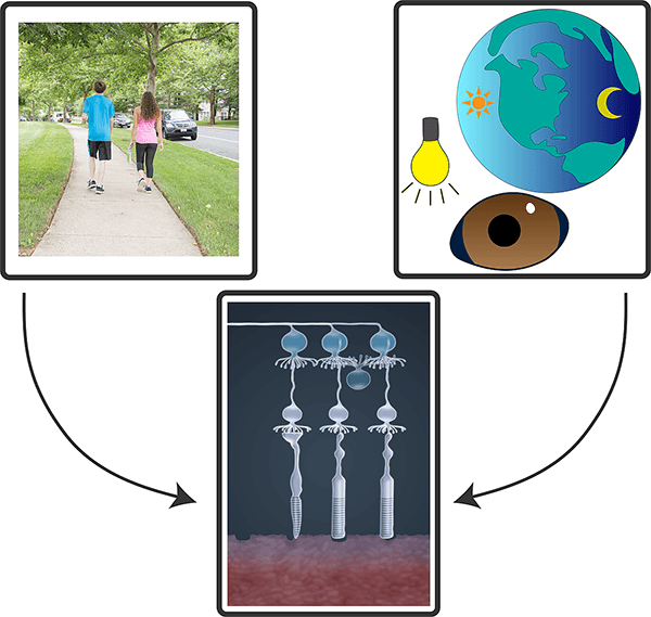
Research advances from the National Institutes of Health (NIH) Intramural Research Program (IRP) often make headlines. Read the news releases that describe our most recent findings:
Loss of the protein pigment epithelium-derived factor (PEDF), which protects retinal support cells, may drive age-related changes in the retina, according to a new study in mice from the National Eye Institute (NEI). The retina is the light-sensitive tissue at the back of the eye, and aging-associated diseases of the retina, like age-related macular degeneration (AMD), can lead to blindness. This new finding could lead to therapies to prevent AMD and other aging conditions of the retina. The study was published in the International Journal of Molecular Sciences. NEI is part of the National Institutes of Health.
“People have called PEDF the ‘youth’ protein, because it is abundant in young retinas, but it declines during aging,” said Patricia Becerra, Ph.D., chief of NEI’s Section of Protein Structure and Function and senior author of the study. “This study showed for the first time that just removing PEDF leads to a host of gene changes that mimic aging in the retina.”
The retina is composed of layers of cells that function together to detect and process light signals, which the brain uses to generate vision. The retina’s light-sensing photoreceptors sit above the retinal pigment epithelium (RPE), a layer of support cells. The RPE nourishes photoreceptors and recycles pieces of the photoreceptor cells called 'outer segments,' which get used up and their tips shed each time photoreceptors detect light. If the RPE cannot provide recycled components of older outer segment tips back to photoreceptors, these cells lose their ability to make new segments, and eventually become unable to sense light. And without nutrients supplied by the RPE, photoreceptors die. In people with AMD or certain types of retinal dystrophies, senescence (aging) or death of RPE cells in the retina leads to vision loss.
RPE from mice without Serpin1 accumulate more lipids than wild-type mice. Super-resolution confocal microscopy of RPE tissue from wild-type (upper) and Serpin1-null (lower) mice. Detailed images on the right are magnified regions of the RPE tissue imaged on the left (dotted square area). RPE cell boundaries are stained in red, and accumulated lipids are stained in green.
Researchers from the National Cancer Institute, part of the National Institutes of Health, and their collaborators have discovered that people of European and African ancestries who were hospitalized for COVID-19 are more likely to carry a particular combination of genetic variants in a gene known as OAS1 than patients with mild disease who were not hospitalized. People with this combination of genetic variants also remain positive for SARS-CoV-2 infection longer. However, interferon treatment may reduce the severity of COVID-19 in people with these genetic factors. Interferons are a type of protein that can help the body’s immune system fight infection and other diseases, such as cancer.
The study appears July 14 in Nature Genetics.
These findings build on previous studies that have suggested that genetic factors, such as genetic variants affecting OAS antiviral proteins that facilitate the detection and breakdown of the SARS-CoV-2 virus, may influence the risk of SARS-CoV-2 infection.
The NCI researchers and their collaborators found that treatment of cells with an interferon decreased the viral load of SARS-CoV-2. The researchers also analyzed data from a clinical trial in which patients with COVID-19 who were not hospitalized were treated with the recombinant interferon pegIFN-λ1 and found that treatment improved viral clearance in all patients; those with the OAS1 risk variants benefitted the most. The results suggest that interferon treatment may improve COVID-19 outcomes and specifically in patients with certain OAS1 genetic variants who have impaired ability to clear infection.
NIH study of pregnant women confirms link with chemicals that could put pregnancy at risk
Pregnant women who were exposed to multiple phthalates during pregnancy had an increased risk of preterm birth, according to new research by the National Institutes of Health. Phthalates are chemicals used in personal care products, such as cosmetics, as well as in solvents, detergents, and food packaging.
After analyzing data from more than 6,000 pregnant women in the United States, researchers found that women with higher concentrations of several phthalate metabolites in their urine were more likely to deliver their babies preterm, which is delivering three or more weeks before a mother’s due date.
“Having a preterm birth can be dangerous for both baby and mom, so it is important to identify risk factors that could prevent it,” said Kelly Ferguson, Ph.D., an epidemiologist at the National Institute of Environmental Health Sciences (NIEHS), part of NIH, and the senior author on the study published in the journal JAMA Pediatrics.
The image shows how a pregnant person may be exposed to phthalates by eating packaged foods and beverages or through personal care product use.
Newly identified brain circuits may point to more effective pain therapies
An international team of scientists has identified the neural mechanisms through which sound blunts pain in mice. The findings, which could inform development of safer methods to treat pain, were published in Science. The study was led by researchers at the National Institute of Dental and Craniofacial Research (NIDCR); the University of Science and Technology of China, Hefei; and Anhui Medical University, Hefei, China. NIDCR is part of the National Institutes of Health.
“We need more effective methods of managing acute and chronic pain, and that starts with gaining a better understanding of the basic neural processes that regulate pain,” said NIDCR Director Rena D’Souza, D.D.S., Ph.D. “By uncovering the circuitry that mediates the pain-reducing effects of sound in mice, this study adds critical knowledge that could ultimately inform new approaches for pain therapy.”
Dating back to 1960, studies in humans have shown that music and other kinds of sound can help alleviate acute and chronic pain, including pain from dental and medical surgery, labor and delivery, and cancer. However, how the brain produces this pain reduction, or analgesia, was less clear.
“Human brain imaging studies have implicated certain areas of the brain in music-induced analgesia, but these are only associations,” said co-senior author Yuanyuan (Kevin) Liu, Ph.D., a Stadtman tenure-track investigator at NIDCR. “In animals, we can more fully explore and manipulate the circuitry to identify the neural substrates involved.”
Sound reduces pain in mice by lowering the activity of neurons in the brain’s auditory cortex (green and magenta) that project to the thalamus.
Photoreceptor cells in mice drive vision and non-vision functions using distinct circuits in the eye
The eye’s light-sensing retina taps different circuits depending on whether it is generating image-forming vision or carrying out a non-vision function such as regulating pupil size or sleep/wake cycles, according to a new mouse study from the National Eye Institute (NEI) and the National Institute of Mental Health (NIMH). The findings could have implications for understanding how our eyes help regulate mood, digestion, sleep, and metabolism. NEI and NIMH are part of the National Institutes of Health.
“We know a lot about pathways involved in image-forming vision, but until now it remained unknown if and how non-image-forming visual behaviors rely on these same pathways in the eye,” said Johan Pahlberg, Ph.D., head of the Photoreceptor Physiology Group at NEI and a senior author of the study.
Vision begins when light travels into the eye and hits the retina’s light-sensing photoreceptors. The photoreceptors transfer signals through several layers of retinal neuron before those signals are sent to the brain. Light also triggers certain non-vision functions, such as controlling how much light enters the eye through the pupil (pupillary light reflex) and regulating the wake/sleep cycle (circadian rhythm). Circadian rhythm disruption has been linked to sleep problems, obesity, and other health issues.
Image-forming and non-image-forming functions use different neuronal circuits in the retina.
Findings could give insight into long-term neurological symptoms of COVID-19
A study from the National Institutes of Health describes the immune response triggered by COVID-19 infection that damages the brain’s blood vessels and may lead to short- and long-term neurological symptoms. In a study published in Brain, researchers from the National Institute of Neurological Disorders and Stroke (NINDS) examined brain changes in nine people who died suddenly after contracting the virus.
The scientists found evidence that antibodies — proteins produced by the immune system in response to viruses and other invaders — are involved in an attack on the cells lining the brain’s blood vessels, leading to inflammation and damage. Consistent with an earlier study from the group, SARS-CoV-2 was not detected in the patients’ brains, suggesting the virus was not infecting the brain directly.
Understanding how SARS-CoV-2 can trigger brain damage may help inform development of therapies for COVID-19 patients who have lingering neurological symptoms.
“Patients often develop neurological complications with COVID-19, but the underlying pathophysiological process is not well understood,” said Avindra Nath, M.D., clinical director at NINDS and the senior author of the study. “We had previously shown blood vessel damage and inflammation in patients’ brains at autopsy, but we didn’t understand the cause of the damage. I think in this paper we’ve gained important insight into the cascade of events.”
SARS-CoV-2 infection can trigger the production of immune molecules that damage cells lining blood vessels in the brain, causing platelets to stick together and form clots. Blood proteins also leak from the blood vessels, leading to inflammation and the destruction of neurons.
A class of viruses known to cause severe diarrheal diseases — including the one famous for widespread outbreaks on cruise ships — can grow in the salivary glands of mice and spread through their saliva, scientists at the National Institutes of Health have discovered. The findings show that a new route of transmission exists for these common viruses, which afflict billions of people each year worldwide and can be deadly.
The transmission of these so-called enteric viruses through saliva suggests that coughing, talking, sneezing, sharing food and utensils, and even kissing all have the potential for spreading the viruses. The new findings still need to be confirmed in human studies.
The findings, which appear in the journal Nature, could lead to better ways to prevent, diagnose, and treat diseases caused by these viruses, potentially saving lives. The study was led by the National Heart, Lung, and Blood Institute (NHLBI), part of NIH.
A microscopic view of salivary gland acinar epithelial cells (pink) infected with rotavirus (green), a type of enteric virus, in a mouse.
Discovery may represent a future target for treating substance use disorders
Researchers have found that blocking certain acetylcholine receptors in the lateral habenula (LHb), an area of the brain that balances reward and aversion, made it harder to resist seeking cocaine in a rat model of impulsive behavior. These findings identify a new role for these receptors that may represent a future target for the development of treatments for cocaine use disorder. There are currently no approved medications to treat cocaine use disorder.
Published in the Journal of Neuroscience, the study was supported by the National Institute on Drug Abuse (NIDA), part of the National Institutes of Health. In 2020, over 41,000 people died from drug overdoses involving stimulants, including cocaine and methamphetamine. Developing safe and effective medications that help treat addictions to cocaine and other stimulants is critical to expand the choices offered to people seeking treatment and to help sustain recovery.
“The LHb acts like an interface between rational thought in the forebrain and the modulation of neurotransmitters like dopamine and serotonin that originate in the midbrain, which are important in regulating decision processes and emotions,” said Carl Lupica, Ph.D., chief of the Electrophysiology Research Section of the Computational and Systems Neuroscience Branch of NIDA. “While the immediate results of this study are related to cocaine seeking, there are also greater implications for impulsivity as it relates to other drugs as well as to psychiatric conditions like obsessive-compulsive disorder. Our future studies will explore the relationship between LHb activity and impulsive behavior related to other drugs such as cannabis, and opioids such as heroin.”
County-level data provides unprecedented detail by geography and population groups
From 2000-2019 overall life expectancy in the United States increased by 2.3 years, but the increase was not consistent among racial and ethnic groups and by geographic area. In addition, most of these gains were prior to 2010. This is according to a new study funded by the National Institutes of Health that examined trends in life expectancy at the county level. The study was led by researchers at the Institute for Health Metrics and Evaluation at the University of Washington’s School of Medicine, Seattle, in collaboration with researchers from NIH and published on June 16th in The Lancet.
“These varied outcomes in life expectancy raise significant questions. Why is life expectancy worse for some and better for others? The novel details in this study provide us the opportunity to evaluate the impact of social and structural determinants on health outcomes in unprecedented ways. This in turn allows us to better identify responsive and enduring interventions for local communities,” said Eliseo J. Pérez-Stable, M.D., co-author and director of the National Institute on Minority Health and Health Disparities (NIMHD), part of NIH.
In most counties, life expectancy for the Black population has increased more than any other racial and ethnic group but overall, the Black population still has a lower life expectancy than the white population. Meanwhile, the white population had a moderate increase, and in some counties, a decrease in life expectancy. Considering these two trends, the study noted that the decrease in the white-Black life expectancy gap could be attributed to the stagnation and reversal of gains in the white population. In addition, American Indian and Alaska Native populations have the lowest life expectancy of all populations and experienced a decrease in most counties, with a gap of more than 21 years in some counties.
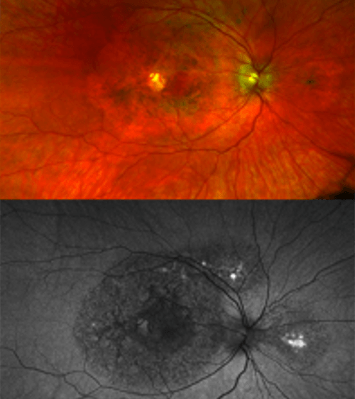
Genetic and clinical research reveals new type of macular dystrophy, a cause of central vision loss
Researchers from the National Eye Institute (NEI) have identified a new disease that affects the macula, a small part of the light-sensing retina needed for sharp, central vision. Scientists report their findings on the novel macular dystrophy, which is yet to be named, in JAMA Ophthalmology. NEI is part of the National Institutes of Health.
Macular dystrophies are disorders that usually cause central visual loss because of mutations in several genes, including ABCA4, BEST1, PRPH2, and TIMP3.
For example, patients with Sorsby Fundus Dystrophy, a genetic eye disease specifically linked to TIMP3 variants, usually develop symptoms in adulthood. They often have sudden changes in visual acuity due to choroidal neovascularization– new, abnormal blood vessels that grow under the retina, leaking fluid and affecting vision.
TIMP3 is a protein that helps regulate retinal blood flow and is secreted from the retinal pigment epithelium (RPE), a layer of tissue that nourishes and supports the retina’s light-sensing photoreceptors. All TIMP3 gene mutations reported are in the mature protein after it has been “cut” from RPE cells in a process called cleavage.
“We found it surprising that two patients had TIMP3 variants not in the mature protein, but in the short signal sequence the gene uses to ‘cut’ the protein from the cells. We showed these variants prevent cleavage, causing the protein to be stuck in the cell, likely leading to retinal pigment epithelium toxicity,” said Bin Guan, Ph.D., lead author.
Retinal images of a patient with a TIMP3 mutation causing atypical symptoms. While there is visible damage in the retina (dark circles), there is no CNV present.