High-Fidelity Stereocilia
Bechara Kachar uses high-tech imaging to understand the cell biology of hearing.

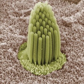
Scanning electron micrograph of the surface epithelium of the frog inner ear
Bechara Kachar’s confocal microscope room could be mistaken for a set from a science fiction movie, with high-tech cabling, lenses, and lasers seemingly strewn in disarray around the room. He describes each instrument proudly, a series of carefully constructed and personally-cared for tools that have helped to make his lab world-famous. “This room is absolutely critical to everything we do—it’s the sole reason we can create the kind of images we produce,” Kachar explains.
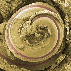
A scanning electron micrograph of the cochlea, a spiral-shaped tunnel in the bony labyrinth of the inner ear
His lab’s high-resolution micrographs of beautifully delicate, staircase-shaped structures of the inner ear, called stereocilia, have graced the covers of many high-profile scientific journals. Stereocilia are miniscule hair-like protrusions on the surface of sensory cells (also called hair cells) found deep within the cochlear and labyrinth structures of the inner ear. They serve as the key mechanosensors, responding to fluid motion for various functions, including hearing and balance. Emphasizing how sensitive these structures are, Kachar describes being able to hear a pin drop from across a room: the sound wave from the pin dropping produces an increase in pressure within the fluid contained in the inner ear, resulting in a shear force that presses the stereocilia against each other. The stereocilia then convert this mechanical movement into electrical signals, which are sent to the brain—all within a matter of milliseconds.
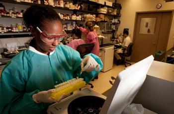
Members of Kachar’s lab investigate fundamental biological processes of hearing
Kachar’s interest in cellular structures and functions began more than three decades ago, but it is the stereocilia’s role in hearing that continues to fascinate him. “Cells in the human body are usually renewed on a regular basis—sometimes at an incredible rate—for example, in the skin and intestines,” says Kachar. “However, this isn’t the case in the inner ear—we’re born with the same cochlear hair cells we die with.” Hair cells gradually develop stereocilia in utero and around birth, after which stereocilia will be with an individual for life: coping with the tantrums of toddlers, the rock concerts of adolescence, and the daily noise of an adult working life. In addition to noise exposure, hair cells and their stereocilia are particularly sensitive to medications that damage the inner ear (ototoxic drugs), such as aspirin, some antibiotics, and some chemotherapy drugs. It is no surprise then that many of us will start to lose our hearing over time.
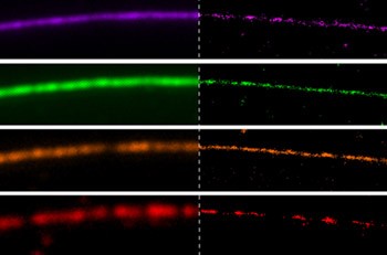
Diffraction-limited microscopy images (Left) and bleaching/blinking assisted localization microscopy (BaLM) images (Right) of a single microtubule labeled with a mixture of fluorescent dyes
To understand gradual hearing loss, Kachar’s team set about investigating how a single cell could maintain such a delicate structure over an entire lifetime. The answer, it seems, may lie in a protein called actin, which underlies many complex structural elements in cells. Kachar’s group discovered a “treadmill-like” motion within neonatal stereocilia where actin molecules constantly build and demolish the internal structure of the protrusion, thus dynamically sculpturing the stereocilia staircase shape. Understanding how that process contributes to long-term maintenance of the stereocilia may provide insights into how to avoid or even repair age-related hearing loss, a translational step into the clinic that will require extensive clinical collaboration.
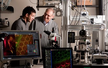
Kachar and his former postdoctoral fellow Uri Manor, Ph.D., discuss microscope design
No stranger to collaborations, Kachar recently partnered with Jennifer Lippincott-Schwartz, Ph.D., Head of the Section on Organelle Biology at the National Institute of Child Health and Human Development (NICHD), to describe a novel microscopy method of viewing individual fluorescently-labeled molecules, and their work was published in the Proceedings of the National Academy of Sciences. He is confident that a similar joint venture would effectively translate his team’s fundamental research into the clinical realm: “In the IRP, you can find a collaborator in the elevator before you go home!” Kachar jokes. But, in all seriousness, he is probably right—and many researchers want to work with him.
In recent years, a stream of fellows and students from top-tier schools have graduated from Kachar’s lab, and, following his lead, many of them are now forging successful careers of their own in related areas of research. On top of pushing the boundaries of science in his lab, Kachar finds it a joy and privilege to mentor young scientists in the early stages of their careers and share with them an intricate symphony composed of complex proteins and delicate structures—the sound of his science.
Bechara Kachar, M.D., is Chief of the Laboratory of Cell Structure and Dynamics at the National Institute on Deafness and Other Communication Disorders (NIDCD).
This page was last updated on Wednesday, May 24, 2023