Better together: Merged microscope offers unprecedented look at biological processes in living cells
NIH researchers combine two microscope technologies to create sharper, faster images
Scientists at the National Institute of Biomedical Imaging and Bioengineering (NIBIB) have combined two different microscope technologies to create sharper images of rapidly moving processes inside a cell. NIBIB is part of the National Institutes of Health.
In a paper published today in Nature Methods, Hari Shroff, Ph.D., chief of NIBIB’s lab section on High Resolution Optical Imaging (HROI), describes his new improvements to traditional Total Internal Reflection Fluorescence (TIRF) microscopy. TIRF microscopy illuminates the sample at a sharp angle so that the light reflects back, illuminating only a thin section of the sample that is extremely close to the coverslip. This process creates very high contrast images because it eliminates much of the background, out-of-focus, light that conventional microscopes pick up.
While TIRF microscopy has been used in cell biology for decades, it produces blurry images of small features within cells. In the past, super-resolution microscopy techniques applied to TIRF microscopes have been able to improve the resolution, but such attempts have always compromised speed, making it impossible to clearly image objects that move rapidly. As a result, many cellular processes remain too small or fast to observe.

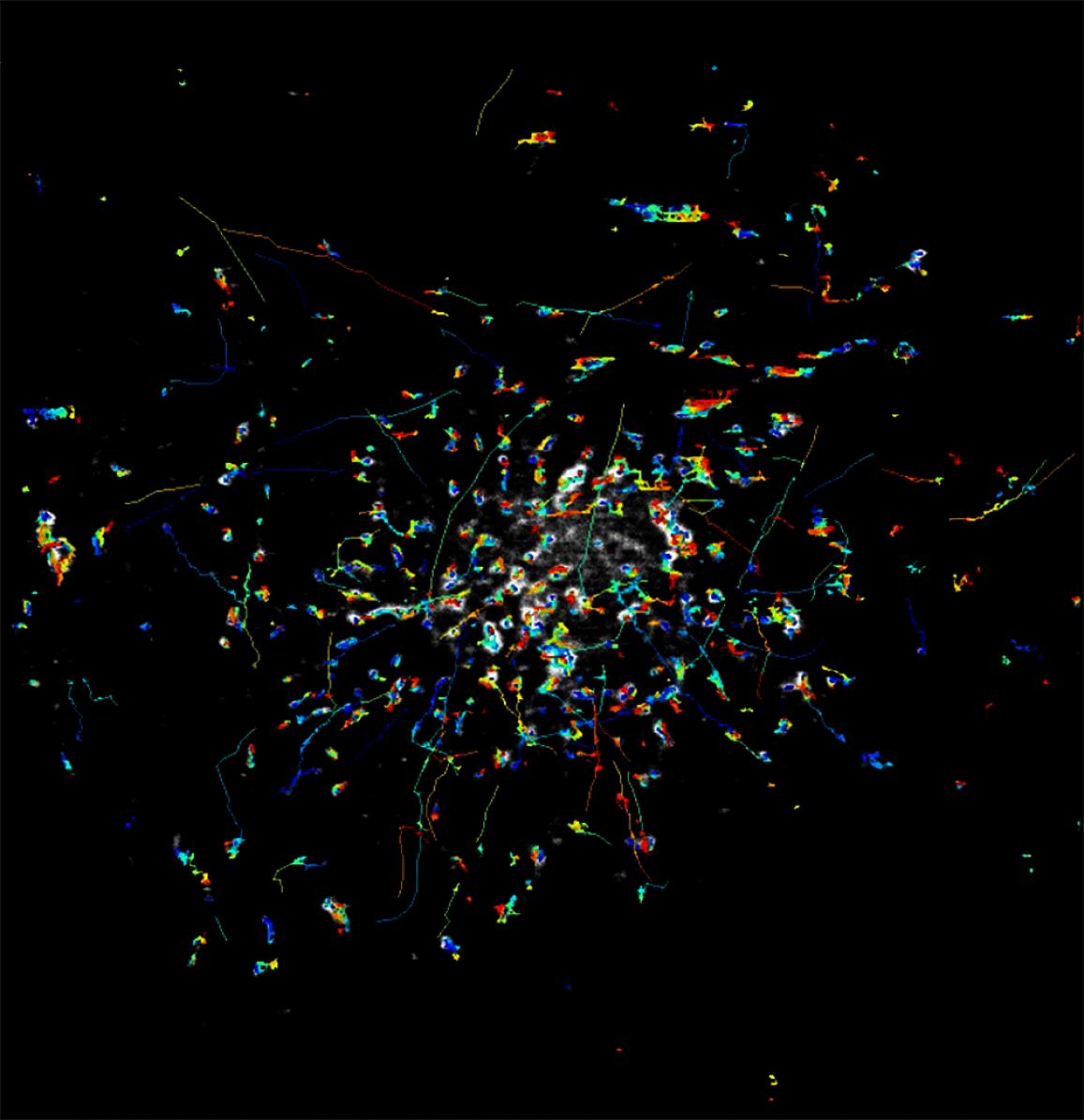
The rapid movements of Rab11 particles can be clearly imaged with the new instant TIRF-SIM microscope.
This page was last updated on Friday, January 21, 2022