Research Topics
Study of miRNA Isoforms (isomiRs)
A specific focus of our research is to elucidate the mechanisms underlying sequence alterations at miRNA ends and how these alterations diversify miRNA function. A single miRNA locus can generate multiple distinct miRNA isoforms (isomiRs) that differ in length, sequence composition, or both. Depending on the site of the heterogeneity, isomiRs can be categorized into 5' and 3' isomiRs. We developed a novel algorithm that allows us to detect and annotate isomiRs from next-generation sequencing data with high confidence. Using this method, we demonstrated that isomiR profiles are cell-, tissue-, and disease-specific. By combining genetic studies with biochemical and structural approaches, we established the tertiary structure of pri-miRNAs and their specific interactions with Drosha as major determinants for 5' isomiR production. Our studies also provided insights into how 5' isomiR biogenesis is regulated in cancer. For 3' isomiRs, we discovered that uridylation can alter the way miRNAs recognize their targets, revealing unique functions of 3' isomiRs. Our findings support the hypothesis that isomiR biogenesis is tightly regulated, allowing isomiRs to play important and distinct biological roles. Current studies are focusing on uncovering novel functions of isomiRs in the context of cancer.
Regulation of miRNAs by Tailing
A unique mode of miRNA regulation is tailing, where non-templated nucleotides are added to the 3' end of pre-miRNA and mature miRNA. By examining the impact of tailing on mature miRNAs, we found that 3' uridylation can alter miRNA target recognition. We also identified the TUT-DIS3L2 machinery as a cellular surveillance system that monitors the 3' ends of AGO-bound miRNAs, with TUT uridylating exposed miRNA 3' ends, leading to degradation by the exonuclease DIS3L2. However, 3' tailing did not globally affect miRNA abundance. In contrast, tailing of pre-miRNAs regulates miRNA abundance through diverse mechanisms. TUT4 and TUT7 negatively regulate let-7 family members by oligo-uridylating their precursors in a LIN28-dependent manner. This oligo-uridylation leads to the degradation of the precursors by the nuclease DIS3L2, resulting in lower levels of mature let-7. Much remains to be understood about the broader impact of oligo-uridylation on miRNA precursors. We aim to understand why certain pre-miRNAs are preferentially uridylated and the functional consequences of pre-miRNA uridylation.
Cancer Mutations in miRNA Biogenesis Pathway Components
Despite the extensive documentation of the roles of miRNAs in tumorigenesis, the development of miRNA-based cancer treatments has lagged. This is partly due to the difficulty in identifying tumor-driver miRNA variations through profiling-based approaches. To address this challenge, we focus on genetic evidence by studying recurrent mutations in miRNA biogenesis factors found in cancer patients. We reasoned that the resulting miRNA alterations are likely crucial to tumor formation and/or progression. Our initial focus is on DICER1 RNase IIIb mutations, which are prominent in certain pediatric cancers and are hallmarks for DICER1 syndrome-related tumors. To elucidate the functional implications of the DICER1 RNase IIIb mutation, we generated a set of cells mimicking DICER1-associated tumors using CRISPR. We found that the DICER1 RNase IIIb hotspot mutation not only causes a loss of function for 5p-miRNAs but also leads to a gain of function for a set of 3p-miRNAs, potentially impacting DICER1 tumor pathogenicity. Our in vitro and in vivo analyses implicate strand switching by AGO as the mechanism for the functional upregulation of passenger 3p-miRNAs, a novel mode of miRNA regulation. Using a mouse model and an isogenic cell culture-based platform, we aim to identify and validate the specific requirements for miRNAs that underlie DICER1-related tumorigenesis. If successful, identifying miRNAs as druggable targets may provide new prospects for cancer therapy.
Biography
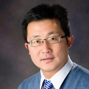
Shuo Gu received his B.A. in Tsinghua University, China. He completed his Ph.D. training in the laboratory of Dr. John Rossi at Beckman Research Institute, City of Hope, Los Angeles. Shuo Gu undertook his postdoctoral training in the laboratory of Dr. Mark Kay at Stanford University Medical School, Palo Alto. Both his Ph.D. and postdoctoral research focused on the mechanisms of RNA interference and microRNA pathways, and their applications in gene therapy. He joined the RNA Biology Laboratory (formerly the Gene Regulation and Chromosome Biology Laboratory) in 2013 as a Stadtman Investigator.
Selected Publications
- Yang A, Bofill-De Ros X, Stanton R, Shao TJ, Villanueva P, Gu S. TENT2, TUT4, and TUT7 selectively regulate miRNA sequence and abundance. Nat Commun. 2022;13(1):5260.
- Yang A, Shao TJ, Bofill-De Ros X, Lian C, Villanueva P, Dai L, Gu S. AGO-bound mature miRNAs are oligouridylated by TUTs and subsequently degraded by DIS3L2. Nat Commun. 2020;11(1):2765.
- Yang A, Bofill-De Ros X, Shao TJ, Jiang M, Li K, Villanueva P, Dai L, Gu S. 3' Uridylation Confers miRNAs with Non-canonical Target Repertoires. Mol Cell. 2019;75(3):511-522.e4.
- Bofill-De Ros X, Kasprzak WK, Bhandari Y, Fan L, Cavanaugh Q, Jiang M, Dai L, Yang A, Shao TJ, Shapiro BA, Wang YX, Gu S. Structural Differences between Pri-miRNA Paralogs Promote Alternative Drosha Cleavage and Expand Target Repertoires. Cell Rep. 2019;26(2):447-459.e4.
- Dai L, Chen K, Youngren B, Kulina J, Yang A, Guo Z, Li J, Yu P, Gu S. Cytoplasmic Drosha activity generated by alternative splicing. Nucleic Acids Res. 2016;44(21):10454-10466.
Related Scientific Focus Areas


Molecular Biology and Biochemistry
View additional Principal Investigators in Molecular Biology and Biochemistry

This page was last updated on Friday, September 20, 2024