Richard D. Leapman, Ph.D.
Senior Investigator
Laboratory of Cellular Imaging and Macromolecular Biophysics
NIBIB
Scientific Director
NIBIB
Research Topics
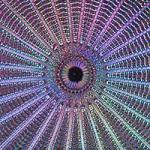
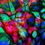
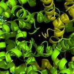
The Section on Cellular and Supramolecular Structure and Function develops new methods based on electron microscopy and related techniques. Our aim is to expand knowledge about complex biological and disease processes, as well as to characterize morphologically the action of diagnostic markers and therapeutic agents in cells. The nanometer scale of biological electron microscopy lies between the realms of live-cell optical microscopy and atomic-scale structural tools of x-ray crystallography and NMR spectroscopy, both of which—unlike EM—require extraction and purification of cellular components. Our approach also complements cryo_EM of isolated frozen-hydrated supramolecular assemblies, which is difficult to apply to intact eukaryotic cells and tissues. We focus on the development of alternative quantitative imaging modes based on detection of elastic and inelastic electron scattering. Current research in the Section Includes development of techniques for (i) determining the tertiary and quaternary structures of macromolecular assemblies, (ii) visualizing 3D ultrastructure, (iii) mapping the elemental composition of subcellular compartments quantitatively, and (iv) studying bionanoparticles and their interactions with cells. We are applying these methods to structural biology, cellular biology, neurobiology, cancer biology, and nanomedicine.
Our group has been particularly active in developing the techniques of electron energy loss spectroscopy (EELS) and combining it with scanning transmission electron microscopy (STEM) to provide an unprecedented high spatial resolution for nanoanalysis of biological structures. We have devised new methods for quantifying both elemental and chemical information obtained from inelastic electron scattering. Recently, our group has extended the techniques of STEM and energy-filtered electron tomography to obtain three-dimensional structural and compositional information about cellular components.
Biography
Dr. Leapman obtained B.A. and M.A. in Natural Sciences, and Ph.D. in physics from the University of Cambridge, UK. He then trained as a postdoctoral fellow in the Department of Materials at the University of Oxford, UK, and in the Department of Applied and Engineering Physics at Cornell University, NY, where he contributed to the development of electron spectroscopy for the nanoscale characterization of materials. He subsequently moved to NIH to develop methods based on scanning transmission electron microscopy and electron spectroscopy to determine the structure and chemical composition of cells and supramolecular assemblies. Dr. Leapman was elected a Fellow of the Microscopy Society of America in 2011. He is currently Editor of the Journal of Microscopy (Oxford), a member of the editorial boards of other microscopy and nanotechnology journals, and has served on numerous national scientific advisory committees. Dr. Leapman is Chief of NIBIB’s Laboratory of Cellular Imaging and Macromolecular Biophysics, while also serving as NIBIB’s Scientific Director since 2006.
Selected Publications
- Aronova MA, Noh SJ, Zhang G, Byrnes C, Meier ER, Kim YC, Leapman RD. Use of dual-electron probes reveals the role of ferritin as an iron depot in ex vivo erythropoiesis. iScience. 2021;24(8):102901.
- Guay MD, Emam ZAS, Anderson AB, Aronova MA, Pokrovskaya ID, Storrie B, Leapman RD. Dense cellular segmentation for EM using 2D-3D neural network ensembles. Sci Rep. 2021;11(1):2561.
- Rao A, McBride EL, Zhang G, Xu H, Cai T, Notkins AL, Aronova MA, Leapman RD. Determination of secretory granule maturation times in pancreatic islet β-cells by serial block-face electron microscopy. J Struct Biol. 2020;212(1):107584.
- He Q, Hsueh M, Zhang G, Joy DC, Leapman RD. Biological serial block face scanning electron microscopy at improved z-resolution based on Monte Carlo model. Sci Rep. 2018;8(1):12985.
- Hohmann-Marriott MF, Sousa AA, Azari AA, Glushakova S, Zhang G, Zimmerberg J, Leapman RD. Nanoscale 3D cellular imaging by axial scanning transmission electron tomography. Nat Methods. 2009;6(10):729-31.
Related Scientific Focus Areas
Biomedical Engineering and Biophysics
View additional Principal Investigators in Biomedical Engineering and Biophysics
This page was last updated on Tuesday, September 7, 2021