Study charts developmental map of inner ear sound sensor in mice
Data offers valuable resource for developing stem cell-based therapies for hearing loss
A team of researchers has generated a developmental map of a key sound-sensing structure in the mouse inner ear. Scientists at the National Institute on Deafness and Other Communication Disorders (NIDCD), part of the National Institutes of Health, and their collaborators analyzed data from 30,000 cells from mouse cochlea, the snail-shaped structure of the inner ear. The results provide insights into the genetic programs that drive the formation of cells important for detecting sounds. The study also sheds light specifically on the underlying cause of hearing loss linked to Ehlers-Danlos syndrome and Loeys-Dietz syndrome.
The study data is shared on a unique platform open to any researcher, creating an unprecedented resource that could catalyze future research on hearing loss. Led by Matthew W. Kelley, Ph.D., chief of the Section on Developmental Neuroscience at the NIDCD, the study appeared online in Nature Communications. The research team includes investigators at the University of Maryland School of Medicine, Baltimore; Decibel Therapeutics, Boston; and King’s College London.
“Unlike many other types of cells in the body, the sensory cells that enable us to hear do not have the capacity to regenerate when they become damaged or diseased,” said NIDCD Director Debara L. Tucci, M.D., who is also an otolaryngology-head and neck surgeon. “By clarifying our understanding of how these cells are formed in the developing inner ear, this work is an important asset for scientists working on stem cell-based therapeutics that may treat or reverse some forms of inner ear hearing loss.”

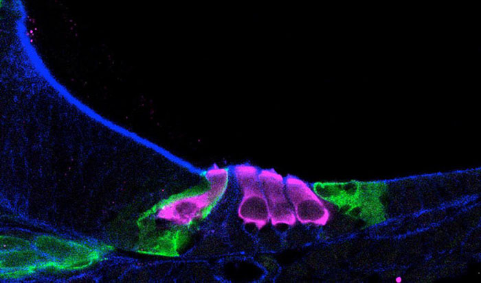
Single-cell RNA sequencing helped scientists map how sensory hair cells (pink) develop in a newborn mouse cochlea.
This page was last updated on Friday, January 21, 2022