Scientists unravel the function of a sight-saving growth factor
NIH study breaks down pigment epithelium-derived factor to understand how it protects and stimulates retinal neurons
Researchers at the National Eye Institute (NEI) have determined how certain short protein fragments, called peptides, can protect neuronal cells found in the light-sensing retina layer at the back of the eye. The peptides might someday be used to treat degenerative retinal diseases, such as age-related macular degeneration (AMD). The study published today in the Journal of Neurochemistry. NEI is part of the National Institutes of Health.
A team led by Patricia Becerra, Ph.D., chief of the NEI Section on Protein Structure and Function, had previously derived these peptides from a protein called pigment epithelium-derived factor (PEDF), which is produced by retinal pigment epithelial cells that line the back of the eye.
“In the eye, PEDF protects neurons from dying. It prevents the invasion of blood vessels, it prevents inflammation, it has antioxidant properties— all these are beneficial properties,” said Becerra, the senior author of the study. Her studies suggest that PEDF is part of the eye’s natural mechanism for maintaining eye health. “PEDF may have a role for treating eye disease. If we want to exploit the protein for therapeutics, we need to separate out the regions responsible for its various properties and determine how each of them works.”

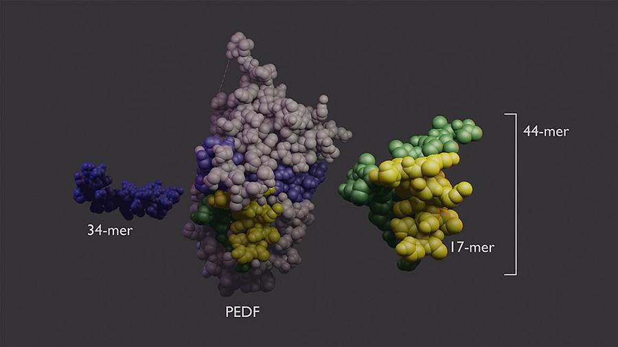
PEDF protein (center) has two domains with different functions. The 34-mer (blue, left) has anti-angiogenic properties. The 44-mer (green and yellow, right) protects and stimulates neurons. The 17-mer (yellow) is a smaller region of the 44-mer with the same function.
This page was last updated on Friday, January 21, 2022