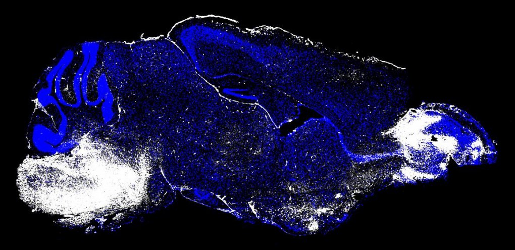
Raising the curtain on cerebral malaria’s deadly agents
NIH scientists film inside mouse brains to uncover biology behind the disease.
Using state-of-the-art brain imaging technology, scientists at the National Institutes of Health filmed what happens in the brains of mice that developed cerebral malaria (CM). The results, published in PLOS Pathogens, reveal the processes that lead to fatal outcomes of the disease and suggest an antibody therapy that may treat it.
This page was last updated on Friday, January 21, 2022
