New Light Microscope Can View Protein Arrangement in Cell Structures
Researchers at Howard Hughes Medical Institute’s Janelia Farm Research Campus, the National Institutes of Health, and Florida State University have developed and applied a new light microscopy technique that will allow them to determine the arrangement of proteins that make up the individual organelles, or structures, within a cell.
The microscope and the technology that make it possible are described in an article appearing on-line in the August 10, 2006, issue of Science Express. The technique was conceived by Eric Betzig, Ph.D., and Harald Hess, Ph.D. while working as independent inventors and later as investigators at Janelia Farm, which subsequently supported their effort on the project. Funding for the project was also provided by the NIH. Drs. Betzig and Hess built the microscope and demonstrated the method at the NIH, while working with Jennifer Lippincott-Schwartz, Ph.D. and her colleagues in the Cell Biology and Metabolism Branch of the National Institute of Child Health and Human Development. Also working on the project was Michael Davidson of the National High Magnetic Field Laboratory at Florida State University.

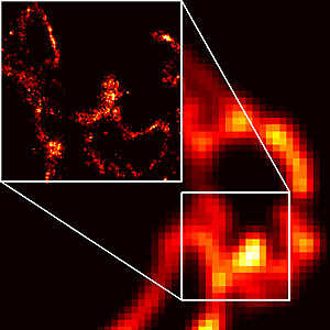
The images depict a membrane protein in a cellular organelle known as a lysosome. The image on the right shows a convention fluorescent image of a portion of the lyososome, whereas the image on the left shows the corresponding PALM image in the region outlined.
This page was last updated on Friday, January 21, 2022