IRP study in mice identifies type of brain cell involved in stuttering
Discovery could lead to targets for new therapies
Researchers believe that stuttering — a potentially lifelong and debilitating speech disorder — stems from problems with the circuits in the brain that control speech, but precisely how and where these problems occur is unknown. Using a mouse model of stuttering, scientists report that a loss of cells in the brain called astrocytes are associated with stuttering. The mice had been engineered with a human gene mutation previously linked to stuttering. The study, which appeared online in the Proceedings of the National Academy of Sciences, offers insights into the neurological deficits associated with stuttering.
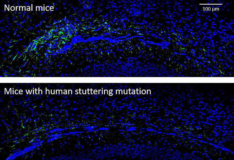
The loss of astrocytes, a supporting cell in the brain, was most prominent in the corpus callosum, a part of the brain that bridges the two hemispheres. Previous imaging studies have identified differences in the brains of people who stutter compared to those who do not. Furthermore, some of these studies in people have revealed structural and functional problems in the same brain region as the new mouse study.
The study was led by Dennis Drayna, Ph.D., of the Section on Genetics of Communication Disorders, at the National Institute on Deafness and Other Communication Disorders (NIDCD), part of the National Institutes of Health. Researchers at the Washington University School of Medicine in St. Louis and from NIH’s National Institute of Biomedical Imaging and Bioengineering, and National Institute of Mental Health collaborated on the research.
“The identification of genetic, molecular, and cellular changes that underlie stuttering has led us to understand persistent stuttering as a brain disorder,” said Andrew Griffith, M.D., Ph.D., NIDCD scientific director. “Perhaps even more importantly, pinpointing the brain region and cells that are involved opens opportunities for novel interventions for stuttering — and possibly other speech disorders.”

In a mouse model of stuttering (lower panel), there are fewer astrocytes, shown in green, compared to controls (upper panel) in the corpus callosum, the area of the brain that enables the left and right hemispheres to communicate.
This page was last updated on Friday, January 21, 2022