IRP researchers decode retinal circuits for circadian rhythm, pupillary light response
Photoreceptor cells in mice drive vision and non-vision functions using distinct circuits in the eye
The eye’s light-sensing retina taps different circuits depending on whether it is generating image-forming vision or carrying out a non-vision function such as regulating pupil size or sleep/wake cycles, according to a new mouse study from the National Eye Institute (NEI) and the National Institute of Mental Health (NIMH). The findings could have implications for understanding how our eyes help regulate mood, digestion, sleep, and metabolism. NEI and NIMH are part of the National Institutes of Health.
“We know a lot about pathways involved in image-forming vision, but until now it remained unknown if and how non-image-forming visual behaviors rely on these same pathways in the eye,” said Johan Pahlberg, Ph.D., head of the Photoreceptor Physiology Group at NEI and a senior author of the study.
Vision begins when light travels into the eye and hits the retina’s light-sensing photoreceptors. The photoreceptors transfer signals through several layers of retinal neuron before those signals are sent to the brain. Light also triggers certain non-vision functions, such as controlling how much light enters the eye through the pupil (pupillary light reflex) and regulating the wake/sleep cycle (circadian rhythm). Circadian rhythm disruption has been linked to sleep problems, obesity, and other health issues.
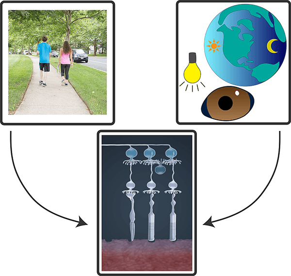
Image-forming and non-image-forming functions use different neuronal circuits in the retina.
This page was last updated on Wednesday, July 27, 2022