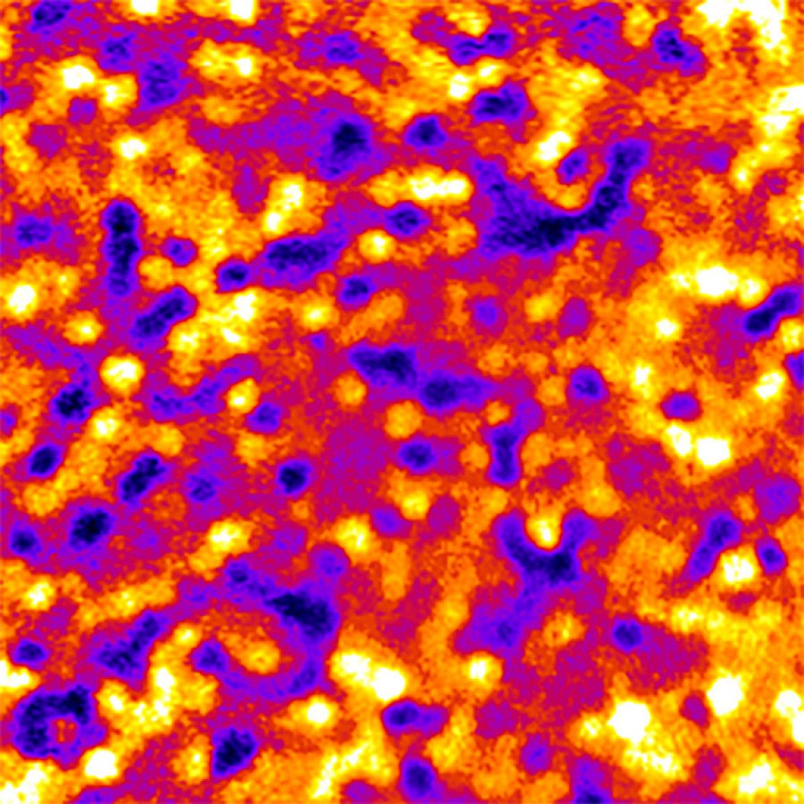
Imaging method reveals long-lived patterns in cells of the eye
Technique could aid early detection and treatment of certain eye diseases
Cells of the retinal pigment epithelium (RPE) form unique patterns that can be used to track changes in this important layer of tissue in the back of the eye, researchers at the National Eye Institute (NEI) have found. Using a combination of adaptive optics imaging and a fluorescent dye, the researchers used the RPE patterns to track individual cells in healthy volunteers and people with retinal disease. The new finding could provide a way to study the progression and treatment of blinding diseases that affect the RPE. The study was published today in the journal JCI Insight. NEI is part of the National Institutes of Health (NIH).
“Studying cells of the retinal pigment epithelium in the clinic is like looking into a black box. RPE cells are difficult to see, and by the time signs of disease are detectable with conventional techniques, a lot of damage has often already occurred,” said Johnny Tam, Ph.D., the lead author of the study. “This study is proof-of-concept that we can use a fluorescent dye to reveal this unique fingerprint of the RPE, and to monitor the tissue over time.”
This page was last updated on Friday, January 21, 2022
