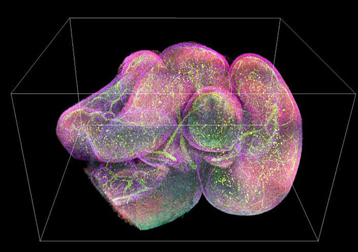
Faster processing makes cutting-edge fluorescence microscopy more accessible
NIH researchers put complex, high-resolution data within reach for many more scientists
Scientists have developed new image processing techniques for microscopes that can reduce post-processing time up to several thousand-fold. The researchers are from the National Institutes of Health with collaborators at the University of Chicago and Zhejiang University, China.
In a paper published in Nature Biotechnology, Hari Shroff, Ph.D., chief of laboratory on High Resolution Optical Imaging at the National Institute of Biomedical Imaging and Bioengineering (NIBIB), describes new techniques that can significantly reduce the time needed to process the highly complex images that are created by the most cutting-edge microscopes. Such microscopes are often used to capture blood and brain cells moving through fish, visualize the neural development of worm embryos, and pinpoint individual organelles within entire organs.
As microscopes continue to get better, creating higher resolution images faster, researchers are finding they have more data than time to process it. While the videos themselves can be captured in minutes, the images could be terabytes in size and require weeks or, in some cases, months of processing time to be useable.
This page was last updated on Friday, January 21, 2022
