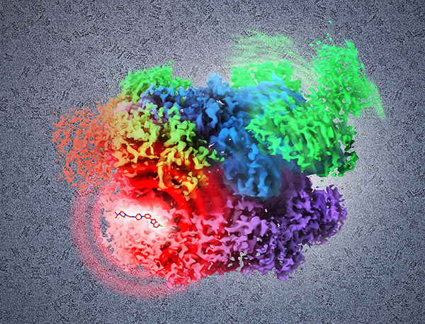
New study using cryo-electron microscopy shows how potential drugs could inhibit cancer
A new study shows that it is possible to use an imaging technique called cryo-electron microscopy (cryo-EM) to view, in atomic detail, the binding of a potential small molecule drug to a key protein in cancer cells. The cryo-EM images also helped the researchers establish, at atomic resolution, the sequence of structural changes that normally occur in the protein, p97, an enzyme critical for protein regulation that is thought to be a novel anti-cancer target.
The study appeared online January 28, 2016, in Science. Sriram Subramaniam, Ph.D., of the National Cancer Institute’s (NCI) Center for Cancer Research, led the research. NCI is part of the National Institutes of Health.
This page was last updated on Friday, January 21, 2022
