IRP researchers develop MRI with lower magnetic field for cardiac and lung imaging
Redesigned MRI holds promise for the diagnosis and treatment of diseases
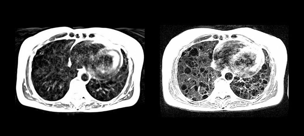
National Institutes of Health researchers, along with researchers at Siemens, have developed a high-performance, low magnetic-field MRI system that vastly improves image quality of the lungs and other internal structures of the human body. The new system is more compatible with interventional devices that could greatly enhance image-guided procedures that diagnose and treat disease, and the system makes medical imaging more affordable and accessible for patients.
The low-field MRI system may also be safer for patients with pacemakers or defibrillators, quieter, and easier to maintain and install. The study, funded by the National Heart, Lung, and Blood Institute (NHLBI), part of the National Institutes of Health, appears today in the journal Radiology.
The trend in recent years has been to develop MRI systems with higher magnetic field strengths to produce clearer images of the brain. But, researchers calculated that using those same state-of-the-art systems — at a modified strength — might offer high quality imaging of the heart and lungs. They found that metal devices such as interventional cardiology tools that were once at risk of heating with the high-field system were now safe for real-time, image-guided procedures such as heart catherization.
“We continue to explore how MRI can be optimized for diagnostic and therapeutic applications,” said Robert Balaban, Ph.D., scientific director of the Division of Intramural Research and chief of the Laboratory of Cardiac Energetics at NHLBI. “The system reduces the risk of heating — a major barrier to the use of MRI-guided therapeutic approaches that have hampered the imaging field for decades.”

Lung cysts and surrounding tissues in a patient with lymphangioleiomyomatosis (LAM) seen more clearly using high-performance low field MRI compared to standard MRI. Photo credit: Campbell-Washburn A E, Ramasawmy R, Restivo M C, et al. Used by permission.
This page was last updated on Friday, January 21, 2022