Research advances from the National Institutes of Health (NIH) Intramural Research Program (IRP) often make headlines. Read the news releases that describe our most recent findings:
Whether you’re shedding pounds with the help of effective new medicines, slimming down after weight loss surgery or cutting calories and adding exercise, there will come a day when the numbers on the scale stop going down, and you hit the dreaded weight loss plateau.
In a recent study, Kevin Hall, a researcher at the National Institutes of Health who specializes in measuring metabolism and weight change, looked at when weight loss typically stops depending on the method people were using todrop pounds. He broke down the plateau into mathematical models using data from high-quality clinical trials of different ways to lose weight to understand why people stop losing when they do. The study published Monday in the journal Obesity.
Newly discovered gene helps some yeast endure toxins and can help scientists understand toxin resistance
National Institutes of Health researchers have identified a gene that makes yeast resistant to a lethal toxin, according to a new study published in the Proceedings of the National Academy of Sciences. To study the evolution of toxin resistance, researchers at the National Human Genome Research Institute (NHGRI), part of NIH, used yeast — the kind commonly used for home baking — as a model organism. While researchers have long known about yeast’s remarkable ability to evade the effects of lethal toxins, the reason was a mystery until now.
“The intricacies of genomics that mediate these within-species battles are beautifully revealed by a study like this,” said Charles Rotimi, Ph.D., scientific director of the Intramural Research Program at NHGRI. “While this is a yeast story, the mechanisms will surely influence studies on toxins and their effects on humans.”
Throughout human history, people have combatted various toxins made by other organisms, like spiders, plants, snakes and even the cholera or anthrax bacteria. Understanding toxin resistance in yeast could lead to new avenues for protection against toxins in humans.
“We’re interested in understanding how genomic variation leads to differences between individuals, so in this study, we’re looking at the most basic biological mechanisms underlying resistance to toxins in simple organisms, such as yeast,” said Meru Sadhu, Ph.D., an investigator in the Genetic Disease Research Branch at NHGRI and senior author of the study. “An important way that organisms vary is in how much they’re affected by toxins.”
NIH researchers discover a possible cause for a rare facial malformation, bringing new hope for patients
Researchers at the National Institutes of Health and their colleagues have found that a toxic protein made by the body called DUX4 may be the cause of two very different rare genetic disorders. For patients who have facioscapulohumeral muscular dystrophy (FSHD), or a rare facial malformation called arhinia, this research discovery may eventually lead to therapies that can help people with these rare diseases.
FSHD type 2 (FSHD2) is an inherited form of muscular dystrophy that causes progressive muscle weakness. Arhinia is an extremely rare yet severe disorder that prevents the development of an external nose and the olfactory bulbs and tracts. Both diseases are caused by mutations in the SMCHD1 gene. In patients with FSHD2, there is overproduction of DUX4 which kills the muscle cells, and this leads to the progressive weakening of the muscles.
“It has been known for some time that DUX4 damages the muscle in patients with FSHD2, but what we found is that it can actually also kill the precursors of the human nose,” said Natalie Shaw, M.D., head of the Pediatric Neuroendocrinology Group at the National Institute of Environmental Health Sciences (NIEHS) and lead author of the new study in the journal Science Advances. NIEHS is part of NIH.
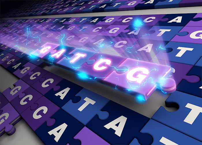
Software opens the door for a greater number of complete genome sequences
National Institutes of Health researchers have developed and released an innovative software tool to assemble truly complete (i.e., gapless) genome sequences from a variety of species. This software, called Verkko, which means 'network' in Finnish, makes the process of assembling complete genome sequences more affordable and accessible. A description of the new software was published today in Nature Biotechnology.
“We took everything we learned in the T2T project and automated the process,” said NHGRI associate investigator Sergey Koren, Ph.D., who led the creation of Verkko and is senior author on the paper. “Now with Verkko, we can essentially push a button and automatically get a complete genome sequence.”
National Institutes of Health researchers have developed and released an innovative software tool to assemble truly complete (i.e., gapless) genome sequences from a variety of species.
Scientists from the National Institute of Allergy and Infectious Diseases (NIAID), part of the National Institutes of Health have removed a major roadblock to better understanding of mpox (formerly, monkeypox). They developed a mouse model of the disease and used it to demonstrate clear differences in virulence among the major genetic groups (clades) of mpox virus (MPXV). The research, appearing in Proceedings of the National Academy of Science, was led by Bernard Moss, M.D., Ph.D., chief of the Genetic Engineering Section of NIAID’s Laboratory of Viral Diseases.
Historically, mpox, a disease resembling smallpox, was only occasionally transmitted from rodents to non-human primates or people and was observed primarily in several African countries. Mpox rarely spread from person to person. That pattern changed in 2022 with an outbreak in which person-to-person mpox transmission occurred in more than 100 locations worldwide. To date, more than 80,000 cases of mpox have been diagnosed during this outbreak. Genome sequencing revealed that the strain causing the current outbreak, clade IIb, differs from two historic clades; clade I, which has a mortality rate of up to 10%, and clade IIa, which has a mortality rate of less than 1%. Mortality from Clade IIb MPXV is lower than either of the historic clades
Colorized scanning electron micrograph of monkeypox virus (blue) on the surface of infected VERO E6 cells (pink).
New disease could provide insights into how the cell’s recycling system contributes to a healthy brain
Researchers at the National Institutes of Health have discovered a new neurological condition characterized by issues with motor coordination and speech. They report their findings in npj Genomic Medicine.
Scientists from NIH’s National Human Genome Research Institute (NHGRI) and Undiagnosed Diseases Program (UDP) identified three children with the condition, two siblings and an unrelated child. The three children all had issues with motor coordination and speech, and one child had abnormalities in the cerebellum, the part of the brain involved in complex movement among other functions. Additionally, the children all had mutations in both copies of the ATG4D gene.
ATG4D aids in the cellular housekeeping process called autophagy, which cells use to break down and recycle damaged proteins and other defective pieces of the cell to stay healthy. Autophagy is a fundamental process used by cells throughout the body, but neurons are particularly dependent on autophagy for survival. However, little is known about how ATG4D contributes to healthy neurons.
“Among genetic diseases, we’ve solved many of the lower hanging fruits,” said May Christine Malicdan, M.D., Ph.D., NHGRI staff scientist and senior author of the study. “Now, we’re reaching for the higher fruits — genes like ATG4D that are more difficult to analyze — and we have the genomic and cellular tools to do so.”
NIH study identifies new molecules involved in diabetes
In a new large-scale genetic analysis, scientists have found a set of small RNA molecules, called microRNAs, in human pancreatic cells that are strongly associated with type 2 diabetes. Researchers discovered the microRNAs in groups of cells called pancreatic islets, which produce hormones, such as insulin, that the body uses to regulate energy levels.
In people with diabetes, the islets fail to produce sufficient insulin to control blood sugar, which is why understanding the basic biology of pancreatic islets is important for human health.
The study, led in part by scientists at the National Human Genome Research Institute (NHGRI), part of the National Institutes of Health, will inform future studies on the early detection and treatment of diabetes. The results were published in Proceedings of the National Academy of the Sciences.
“This study represents the largest sequenced-based analysis of microRNA expression in human pancreatic islets to date,” said Francis Collins, M.D., Ph.D., senior investigator in the Center for Precision Health Research in NHGRI’s Intramural Research Program and senior author of the study. “The results of this study set the stage for understanding how microRNAs fine-tune gene expression in pancreatic islets and its implications for diabetes.”
New inoculation based on Ebola VSV vaccine concept
A National Institutes of Health research group with extensive experience studying ebolavirus countermeasures has successfully developed a vaccine against Sudan virus (SUDV) based on the licensed Ebola virus (EBOV) vaccine. SUDV, identified in 1976, is one of the four viruses known to cause human Ebolavirus disease. The new vaccine, VSV-SUDV, completely protected cynomolgus macaques against a lethal SUDV challenge. The findings were published in the journal The Lancet Microbe.
SUDV is distinct from and less common than EBOV, but similarly deadly. A recent four-month SUDV outbreak in Uganda that ended on Jan. 11, 2023, caused 142 confirmed cases and 55 deaths. No treatment or vaccine for SUDV disease is licensed, although candidates are in clinical and preclinical trials. One of these candidates is VSV-SUDV, developed and tested by scientists at NIH’s National Institute of Allergy and Infectious Diseases in Hamilton, Montana.
The live-attenuated vaccine uses genetically engineered vesicular stomatitis virus (VSV), an animal virus that primarily affects cattle, to express a SUDV protein as a single-dose vaccine. The researchers developed VSV-SUDV using techniques that led to Ervebo, the VSV-EBOV vaccine that the European Medicines Agency and the U.S. Food and Drug Administration approved in 2019 as the first vaccine for the prevention of Ebola virus disease. In the current studies, the investigators replaced the key EBOV protein in Ervebo with the comparable protein from SUDV.
Colorized scanning electron micrograph of filamentous Ebola virus particles (green) attached to and budding from a chronically infected VERO E6 cell (blue).
New research offers clues about the biology of cells in the spinal cord that die off in amyotrophic lateral sclerosis (ALS) and other neurodegenerative diseases. A team of researchers supported by the National Institutes of Health found evidence linking motor neurons’ large cell size and supporting structure with the genes that underlie their vulnerability to degeneration in ALS. Findings appeared in Neuron.
The study resulted in a catalog or 'atlas' characterizing the diverse community of cell types within the human spinal cord. By examining gene expression at the single-cell level, the researchers identified dozens of cell types in the spinal cord and analyzed their molecular profiles. They demonstrated the usefulness of the atlas by looking closely at motor neurons, which provide voluntary movement and motor control. Motor neurons, which degenerate and die in ALS, are large cells with one long extension called an axon ― up to a meter long ― that conducts signals from the spinal cord to the muscle fiber.
The team found that motor neurons are distinguished by a set of genes that may enable the large size of the motor neuron cell body and lengthy axon, but also underlie their vulnerability to degeneration. Their molecular profile was defined by genes involved in cytoskeletal structure, which gives the cell shape and organizes the structures within; neurofilament genes related to cell size; and genes linked to the onset of ALS. Additional experiments showed that ALS-related genes are also enriched in motor neurons in mice. Taken together, the study gives insight into ALS and demonstrates the utility of the spinal cell atlas to study disease and possible interventions.
A newly published paper in The Lancet shows that an experimental vaccine against Marburg virus (MARV) was safe and induced an immune response in a small, first-in-human clinical trial. The vaccine, developed by researchers at the National Institute of Allergy and Infectious Diseases (NIAID), part of the National Institutes of Health, could someday be an important tool to respond to Marburg virus outbreaks.
This first-in-human, Phase 1 study tested an experimental MARV vaccine candidate, known as cAd3-Marburg, which was developed at NIAID’s Vaccine Research Center (VRC). This vaccine uses a modified chimpanzee adenovirus called cAd3, which can no longer replicate or infect cells, and displays a glycoprotein found on the surface of MARV to induce immune responses against the virus. The cAd3 vaccine platform demonstrated a good safety profile in prior clinical trials when used in investigational Ebola virus and Sudan virus vaccines developed by the VRC.
Colorized scanning electron micrograph of Marburg virus particles (blue) both budding and attached to the surface of infected VERO E6 cells (orange).
NIH-wide resource will champion diversity, equity, inclusion and accessibility
The National Institute of Biomedical Imaging and Bioengineering (NIBIB) has established the Center for Biomedical Engineering Technology Acceleration (BETA Center), a new intramural research program to solve a range of medicine’s most pressing problems. The BETA Center will serve the wider NIH intramural research program as a biotechnology resource and catalyst for NIH research discoveries.
The center will incorporate a focused engineering approach to accelerate the development, validation and dissemination of cutting-edge technologies. Areas of emphasis will include biomedical imaging, biosensing, engineered and synthetic biology, nanomaterials and biomaterials, artificial intelligence, modeling, computation and informatics. A unique feature of the center will be its ability to rapidly assemble expert teams for purpose-driven technology development to address urgent national and global health needs.
“The BETA Center will be a catalyst for innovation and collaboration,” said NIBIB Director Bruce J. Tromberg, Ph.D. “Engineering and technology development are central to everything that NIH does. New tech drives new biomedical discoveries, and new discoveries are transformed into cutting-edge methods, devices and knowledge that can be widely disseminated.”
A fundamental objective of the BETA Center is to expand diversity, equity, inclusion and accessibility (DEIA) at NIBIB, building on the inherent interdisciplinary nature of biomedical engineering.