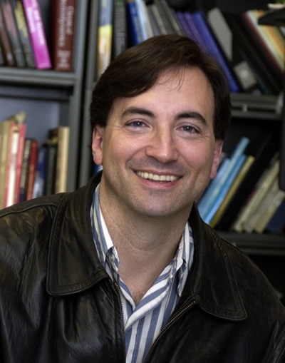
Peter Joel Basser, Ph.D.
Senior Investigator
Section on Quantitative Imaging and Tissue Sciences
NICHD/DIR
Research Topics
Quantitative Imaging and Tissue Sciences
In our basic tissue sciences research, we strive to understand fundamental relationships between function and structure in living tissues. Specifically, we are interested in how microstructure, hierarchical organization, composition, and material and transport properties all affect a tissue's normal functional properties or its dysfunction. Specifically, we view various transport mechanisms as mediating physiological processes. We study these in biological and other model systems over a range of different length and time scales, developing and applying physical/mathematical models (e.g., molecular dynamics and continuum mechanics) to explain tissue functional properties and behavior, and performing physical measurements in tandem. Experimentally, we use water to probe both steady-state and dynamic interactions among tissue constituents from nanometers to centimeters and from nanoseconds to decades. To determine the equilibrium osmo-mechanical properties of well defined model systems, we vary water content or ionic composition systematically. To probe tissue structure and dynamics, we employ atomic force microscopy (AFM), small-angle X-ray scattering (SAXS), small-angle neutron scattering (SANS), static light scattering (SLS), dynamic light scattering (DLS), and one-, two- and multi-dimensional nuclear magnetic resonance (NMR) relaxometry, diffusometry and exchange methods. A goal of our basic tissue sciences research is to develop new understanding and tools that can readily be translated from bench-based quantitative methodologies to the bedside.
Our tissue sciences research above dovetails with our basic and applied research in quantitative imaging, which is intended to generate measurements and maps of intrinsic physical quantities, including diffusivities, relaxivities, or exchange rates, rather than qualitative "stains" and images displayed in a conventional radiological exams. At a basic level, our translational work is directed toward making key 'invisible' structures and processes 'visible'. Our quantitative imaging group uses knowledge of physics, engineering, applied mathematics, imaging and computer sciences, and insights gleaned from our tissue sciences research to discover, develop and test novel quantitative imaging biomarkers that have the potential to sensitively and specifically detect changes in tissue composition, microstructure, or microdynamic processes. The ultimate translational goal of developing such biomarkers is to assess normal and abnormal development, diagnose childhood diseases and disorders, and characterize degeneration and trauma. Primarily, we use MRI as the imaging modality of choice because it is so well suited to important NICHD mission–critical applications; it is non-invasive, non-ionizing, requires (in most cases) no exogenous contrast agents or dyes, and is generally deemed safe and effective for fetuses, mothers, and children in both clinical and research settings.
A technical goal of ours has been to transform clinical MRI scanners into scientific instruments capable of producing reproducible, accurate, and precise imaging data to enable the measurement and mapping of useful imaging biomarkers for various biological and clinical applications, including single scans, longitudinal and multi-site studies, personalized medicine, genotype/phenotype correlation studies, and even populating imaging databases with high-quality subject or patient data. With the advent of low-cost, low-field MRI systems, we have begun working to improve image quality, sensitivity and specificity as a means to democratize access to clinical scanning resources.
Biography
Dr. Peter Basser received his A.B., S.M., and Ph.D. degrees in Engineering Sciences from Harvard University and his postdoctoral training in Bioengineering within the NIH IRP. In 1998, he became Chief of the new Section on Tissue Biophysics and Biomimetics (STBB), NICHD and a Senior Investigator. From 2009 through 2015, he additionally served as Director of the Program on Pediatric Imaging and Tissue Sciences (PPITS), and Head of the new Section on Quantitative Imaging and Tissue Sciences, and then was appointed as Associate Scientific Director for Translational Imaging and Genomic Integrity within the NICHD IRP. He currently heads the Affinity Group on Maternal-Fetal Health and Translational Imaging within the NICHD IRP.
Dr. Basser is widely known for the invention, development, and clinical implementation of MR diffusion tensor imaging (DTI), diffusion tensor "streamline tractography," and other quantitative MRI methods for performing in vivo MRI histology or "microstructure imaging". These include CHARMED and AxCaliber MRI, which measures the mean axon diameter and axon diameter distribution, respectively, within white matter pathways, and various double Pulsed-Field Gradient (dPFG) or double wave-vector MRI methods, which are now widely used to elucidate distinct microstructural features of both gray and white matter in the brain. Within the area of neurotechnology, he made seminal contributions to our understanding of the physical underpinnings of transcranial magnetic stimulation (TMS) and even its application to treating depression. He also wrote the first paper describing a new technique for delivering chemotherapeutic agents to the brain, which is now called "convection enhanced delivery" or CED.
Dr. Basser's notable awards and achievements include induction into the National Academy of Engineering (NAE) and the National Academy of Inventors (NAI), and receipt of the Gold Medal from the International Society of Magnetic Resonance in Medicine (ISMRM), and the Eduard Rhein Foundation Technology Award, as well as several awards from national radiological societies.
Selected Publications
- Komlosh ME, Horkay F, Freidlin RZ, Nevo U, Assaf Y, Basser PJ. Detection of microscopic anisotropy in gray matter and in a novel tissue phantom using double Pulsed Gradient Spin Echo MR. J Magn Reson. 2007;189(1):38-45.
- Basser PJ, Pajevic S, Pierpaoli C, Duda J, Aldroubi A. In vivo fiber tractography using DT-MRI data. Magn Reson Med. 2000;44(4):625-32.
- Özarslan E, Koay CG, Shepherd TM, Komlosh ME, İrfanoğlu MO, Pierpaoli C, Basser PJ. Mean apparent propagator (MAP) MRI: a novel diffusion imaging method for mapping tissue microstructure. Neuroimage. 2013;78:16-32.
- Basser PJ, Pajevic S. A normal distribution for tensor-valued random variables: applications to diffusion tensor MRI. IEEE Trans Med Imaging. 2003;22(7):785-94.
- Assaf Y, Blumenfeld-Katzir T, Yovel Y, Basser PJ. AxCaliber: a method for measuring axon diameter distribution from diffusion MRI. Magn Reson Med. 2008;59(6):1347-54.
Related Scientific Focus Areas
Biomedical Engineering and Biophysics
View additional Principal Investigators in Biomedical Engineering and Biophysics
This page was last updated on Monday, December 2, 2024