
Research Topics
Exocytosis, Endocytosis, and Imaging Technology Development
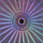
Cells communicate with each other by releasing proteins, peptides, and chemicals through a process called exocytosis–the fusion of cargo-loaded vesicles in the cytoplasm with the plasma membrane. Triggered exocytosis is a highly regulated process. Cells go to great lengths to ensure that exocytosis occurs at precisely the right time and location, and that the correct quantity and type of materials are released during each exocytic burst. Once vesicles fuse, vesicle material is cleared from the plasma membrane through a coat-driven retrieval process called endocytosis. The primary retrieval mechanism in eukaryotic cells is clathrin-mediated endocytosis (CME). In CME, dozens of proteins capture, polymerize, and bend a honeycomb-like coat around pieces of the plasma membrane. How this complex ensemble of protein is organized, however, is largely unknown.
In a healthy cell, these two processes, exocytosis and endocytosis, are carefully balanced. Disruption of either can result in deficiencies or excesses of signals, leading to diseases such as Parkinson’s, schizophrenia, epilepsy, diabetes, and Huntington’s disease. Not surprisingly, understanding exocytosis and endocytosis has been a major goal for the biological sciences. The accumulation of data from many fields, including genetics, biochemistry, cell biology, and structural biology, has provided a spectacular understanding of this biology. Yet much remains to be discovered. Specifically, it is not well understood how dozens of different molecules work together in the crowded cellular environment to regulate and catalyze exocytosis and endocytosis. Furthermore, the central question of how these processes are carefully balanced even under drastic changes in cellular activity is unclear. Thus, the Taraska lab’s long-term goal is to understand the basic molecular foundations of triggered exocytosis and endocytosis. We aim to accomplish this by providing a detailed but comprehensive spatial and temporal molecular map of these processes in living cells.
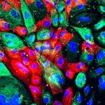
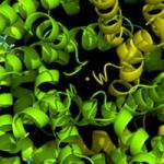
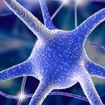
At its core, the Taraska lab develops and uses advanced fluorescence and electron microscopy methods along with structural, biochemical, cellular, and biophysical tools to investigate the nanometer-scale organization of proteins that regulate key cellular events. A gap exists between understanding protein structures and their dynamic cellular contexts. As more atomic structures are solved, filling this gap becomes more important and challenging. We aim to fill this gap by developing and using new ultra-high resolution imaging tools to determine the nano-scale structures, organizations, and dynamics of molecules that are important for the biology of membrane traffic in neurons, endocrine, and immune cells.
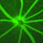
We map the fundamental architecture of molecular machines to understand how these complex assemblies function in living cells.
Biography
Justin Taraska received his B.A. in biology from Reed College in 1999 and earned his Ph.D. in cell biology from Oregon Health and Science University in 2004 in the laboratory of Wolfhard Almers. He conducted his postdoctoral research in the laboratory of William Zagotta at the University of Washington during which time he received a Jane Coffin Child Memorial Fellowship. In 2010, Dr. Taraska became a tenure-track Investigator at the NHLBI. Dr. Taraska is a 2012 PECASE recipient, the highest honor bestowed by the U.S. government on outstanding scientists and engineers beginning their independent careers. Dr. Taraska is a member of the Biophysical Society and the American Society for Cell Biology. He is also faculty in the analytical and quantitative light microscopy course (AQLM) at the Marine Biological Lab in Woods Hole, MA.
Selected Publications
- Obashi K, Sochacki KA, Strub MP, Taraska JW. A conformational switch in clathrin light chain regulates lattice structure and endocytosis at the plasma membrane of mammalian cells. Nat Commun. 2023;14(1):732.
- Alfonzo-Méndez MA, Sochacki KA, Strub MP, Taraska JW. Dual clathrin and integrin signaling systems regulate growth factor receptor activation. Nat Commun. 2022;13(1):905.
- Sochacki KA, Heine BL, Haber GJ, Jimah JR, Prasai B, Alfonzo-Méndez MA, Roberts AD, Somasundaram A, Hinshaw JE, Taraska JW. The structure and spontaneous curvature of clathrin lattices at the plasma membrane. Dev Cell. 2021;56(8):1131-1146.e3.
- Kong L, Sochacki KA, Wang H, Fang S, Canagarajah B, Kehr AD, Rice WJ, Strub MP, Taraska JW, Hinshaw JE. Cryo-EM of the dynamin polymer assembled on lipid membrane. Nature. 2018;560(7717):258-262.
- Sochacki KA, Dickey AM, Strub MP, Taraska JW. Endocytic proteins are partitioned at the edge of the clathrin lattice in mammalian cells. Nat Cell Biol. 2017;19(4):352-361.
Related Scientific Focus Areas
Biomedical Engineering and Biophysics
View additional Principal Investigators in Biomedical Engineering and Biophysics
This page was last updated on Thursday, November 14, 2024