
Jennifer A. Lippincott Schwartz, Ph.D.
Adjunct Investigator
Advanced Microscopy Facility
NICHD/DIR
Research Topics
Dynamics of Membrane Trafficking, Sorting, and Compartmentalization Within Eukaryotic Cells
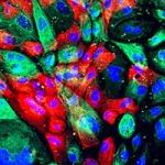
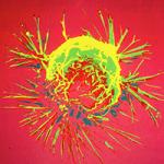
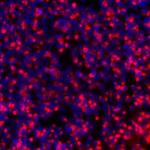
We investigate the global principles underlying secretory membrane trafficking, sorting, and compartmentalization within eukaryotic cells. We use live-cell imaging of green fluorescent protein (GFP) fusion proteins in combination with photobleaching and photoactivation techniques to investigate the subcellular localization, mobility, transport routes, and turnover of a variety of proteins with important roles in the organization and regulation of membrane trafficking and compartmentalization. To test mechanistic hypotheses about protein and organelle dynamics, we use quantitative measurements of these protein characteristics in kinetic modeling and simulation experiments. Among the topics under investigation are (i) membrane partitioning and its role in protein sorting and transport in the Golgi apparatus; (ii) biogenesis and dynamics of unconventional organelles; (iii) mitochondrial morphology and its regulation of cell cycle progression; and (iv) cytoskeletal and endomembrane cross-talk in polarized epithelial cells, hematopoietic niche cells, and the developing Drosophila syncytial blastoderm embryo. We also devoted considerable effort to developing new fluorescence microscopy techniques for imaging fluorescently tagged proteins at near-molecular resolution.
Biography
Dr. Jennifer Lippincott-Schwartz obtained her Ph.D. from Johns Hopkins University in Baltimore, MD and received postdoctoral training with Dr. Richard Klausner of NICHD, NIH. She was Chief of the Section on Organelle Biology in the Cell Biology and Metabolism Branch of the NICHD before becoming a group leader at HHMI's Janelia Research Campus in Ashburn, VA in March 2016. Dr. Lippincott-Schwartz's research uses live cell imaging approaches to analyze the spatio-temporal behavior and dynamic interactions of molecules in cells. These approaches have helped to change the conventional “static” view of protein distribution and function in cells to a more dynamic view that integrates information on protein localization, concentration, diffusion, and interactions that are indiscernible from protein sequences and in vitro biochemical experiments alone. The projects in Lippincott-Schwartz's lab cover a vast range of cell biological topics, including protein transport and the cytoskeleton, organelle assembly and disassembly, and the generation of cell polarity. Analysis of the dynamics of fluorescently labeled proteins expressed in cells is performed using numerous live cell imaging approaches, including FRAP, FCS, and photoactivation. Most recently, her research has focused on the development and use of photoactivatable fluorescent proteins, which “switch on” in response to uv light. One application of these proteins she has put to use is in photoactivated localization microscopy (i.e., PALM), a superresolution imaging technique that enables visualization of molecule distributions at high density at the nano-scale. Dr. Lippincott-Schwartz serves as Editor for Current Protocols in Cell Biology, Molecular Biology of the Cell, and The Journal of Cell Science, and is on the Editorial Board of Cell. She is an active member of the scientific community, having served as President of the American Society of Cell Biology and as Council member of of the Biophysical Society. She was elected to the National Academy of Sciences in 2008 and the National Institute of Medicine in 2009.
Selected Publications
- Satpute-Krishnan P, Ajinkya M, Bhat S, Itakura E, Hegde RS, Lippincott-Schwartz J. ER stress-induced clearance of misfolded GPI-anchored proteins via the secretory pathway. Cell. 2014;158(3):522-33.
- Manor U, Bartholomew S, Golani G, Christenson E, Kozlov M, Higgs H, Spudich J, Lippincott-Schwartz J. A mitochondria-anchored isoform of the actin-nucleating spire protein regulates mitochondrial division. Elife. 2015;4.
- Burnette DT, Shao L, Ott C, Pasapera AM, Fischer RS, Baird MA, Der Loughian C, Delanoe-Ayari H, Paszek MJ, Davidson MW, Betzig E, Lippincott-Schwartz J. A contractile and counterbalancing adhesion system controls the 3D shape of crawling cells. J Cell Biol. 2014;205(1):83-96.
- Van Engelenburg SB, Shtengel G, Sengupta P, Waki K, Jarnik M, Ablan SD, Freed EO, Hess HF, Lippincott-Schwartz J. Distribution of ESCRT machinery at HIV assembly sites reveals virus scaffolding of ESCRT subunits. Science. 2014;343(6171):653-6.
- Rambold AS, Cohen S, Lippincott-Schwartz J. Fatty acid trafficking in starved cells: regulation by lipid droplet lipolysis, autophagy, and mitochondrial fusion dynamics. Dev Cell. 2015;32(6):678-92.
Related Scientific Focus Areas
Biomedical Engineering and Biophysics
View additional Principal Investigators in Biomedical Engineering and Biophysics
This page was last updated on Wednesday, November 8, 2017