
Research Topics
The Molecular Biology Section (MBS) is directed toward a precise molecular understanding of critical interactions that initiate, control, and perpetuate the immune response. To this aim, the laboratory focuses on
- The cell surface molecules encoded by the major histocompatibility complex (MHC), both the MHC class I and class II molecules, the function of which is to present antigens to T lymphocytes
- Efforts are focused on both functional and structural studies, including understanding the molecular mechanisms of peptide loading.
- Viral immunoevasins related to the MHC-I molecules, particularly those encoded by the cytomegaloviruses
- Immune hypersensitivity reactions related to the expression of specific MHC-I molecules, in particular the role of the human MHC-I molecule HLA-B*57:01 in hypersensitivity to the antiretroviral drug, abacavir
- T-lymphocyte receptors (TCR), which by clonal expression confer antigen and MHC specificity for the activation of T lymphocytes
- Natural-killer (NK)-cell receptors, cell surface molecules of effector cells of the innate immune system, that mediate recognition of tumor and virally infected cells via the level and composition of MHC-I molecules on the NK cell target
In recent years, the major accomplishments of MBS have included determination of the structure and function of the CD8 alpha beta T-cell coreceptor in complex with its MHC-I ligand, studies of the mechanism of peptide loading of the MHC-I molecule, and studies of the structure and function of viral immunoevasins. These last advances include the determination of the structure of the complex of the mouse cytomegalovirus MHC-I-like molecule m152 in complex with its ligand RAE1.
Using purified MHC molecules, viral immunoevasins, TCR, and NK receptors, we have learned the thresholds of affinity that allow such cell surface receptors to activate or inhibit effector-cell function. Based on X-ray crystallographic studies of these components and of their molecular complexes, we now understand in molecular detail the mechanism of peptide loading of MHC-I molecules, the basis of molecular recognition of TCR for MHC-I and MHC-II molecules, and the structural basis for interaction of T-cell coreceptors with MHC molecules. X-ray structures of MHC-II/peptide complexes that are crucial to the development of autoimmunity provide insight into the molecular basis of autoimmunity.
Our recent studies of viral immunoevasins reveal the structural evolution of viral resistance to recognition by the immune system and provide opportunities for developing strategies to counter such viral evasion. In addition, our broad understanding of the structure-function relationships of MHC molecules has permitted us to explore the relationship of MHC-I structure to HLA-linked drug-induced hypersensitivity syndromes. We are currently developing mouse model systems to allow further exploration of the molecular and cellular basis of such HLA-linked syndromes.
Credit: NIAIDFigure 1. Transition of the 3,10 helix of the MHC-I molecule H2-Ld upon peptide loading. As described (Mage et al, 2012, 2013), the MHC-I molecule undergoes a structural transition upon peptide binding in the endoplasmic reticulum.Figure 1. Transition of the 3,10 helix of the MHC-I molecule H2-Ld upon peptide loading. As described (Mageet al, 2012, 2013), the MHC-I molecule undergoes a structural transition upon peptide binding in the endoplasmic reticulum.
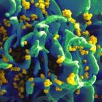
Credit: NIAIDFigure 2. Structure of the molecular complex of the mouse cytomegalovirus protein, m152 (magenta) with its stress-induced ligand, RAE1gamma (green). As described (Wang et al, 2012), the viral immunoevasin controls the surface expression of the stress-induFigure 2. Structure of the molecular complex of the mouse cytomegalovirus protein, m152 (magenta) with its stress-induced ligand, RAE1gamma (green). As described (Wang et al, 2012), the viral immunoevasin controls the surface expression of the stress-induced ligand by binding RAE1 via a pincer-like mechanism.
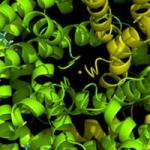
Credit: NIAIDFigure 3. Structure of the complex of the CD8 alpha/beta coreceptor with its MHC-I ligand. As published (Wang et al, 2009), the MHC-I molecule, H2-2Dd (heavy chain, green, light chain, cyan, peptide, tan), is engaged by the CD8 alpha/beta heterodimer (CD8Figure 3. Structure of the complex of the CD8 alpha/beta coreceptor with its MHC-I ligand. As published (Wang et al, 2009), the MHC-I molecule, H2-2Dd (heavy chain, green, light chain, cyan, peptide, tan), is engaged by the CD8 alpha/beta heterodimer (CD8 alpha, magenta; beta, yellow) through interactions with the membrane proximal alpha3 domain of the H2-Dd molecule.
Biography
Biography
Dr. Margulies received an A.B. from Columbia University in 1971. In 1978, he earned his M.D. and Ph.D. from the Albert Einstein College of Medicine. From 1978 to 1980, he served as a resident in medicine at Columbia/Presbyterian Medical Center. From 1980 to 1983, he worked as a research associate in the Laboratory of Molecular Genetics at the National Institute of Child Health and Human Development. From 1983 to 1987, he was an investigator in the Laboratory of Immunology. In 1987, he became a senior investigator and, since 1989, has been chief of the Molecular Biology Section. Since 2008, he has been a member of the Senior Biomedical Research Service.
Awards
Phi Beta Kappa; U.S. Public Health Service Commendation Medal, 1987; Outstanding Service Medal, 1991; Meritorious Service Medal, 1997; Distinguished Service Medal, 2001; NIH Merit Award, 2009 (Tetramer Facility Team)
Memberships
- The American Association of Immunologists
- American Society for Clinical Investigation
- American Association for the Advancement of Science
- Federation of American Scientists
- Protein Society
Advisory/Review Committees
- Immunology and Immunotherapy Advisory Committee (American Cancer Society, 1988-1991)
- Fellowship Subcommittee (Arthritis Foundation, 1989-1991)
- NIH Research Scholars Program Committee (Howard Hughes Medical Institute, 1991-1993)
- National Multiple Sclerosis Society Advisory Committee, 1993-1999
- The American Association of Immunologists, Publications Committee, 1995-1999
- Department of Veterans’ Affairs Joint Biomedical Development Scientific Merit Review Board, Subcommittee for Immunology-B, 2005-2009
- NIH Immunology Interest Group Steering Committee, 1997-1998, 2013-2014
Editorial Boards
- The Journal of Immunology (section editor, 1993-1998)
- Current Protocols in Immunology (1989-date)
- Molecular Immunology (2002-date)
Special Interest Groups
Immunology, Structural Biology
Selected Publications
- Jiang J, Taylor DK, Kim EJ, Boyd LF, Ahmad J, Mage MG, Truong HV, Woodward CH, Sgourakis NG, Cresswell P, Margulies DH, Natarajan K. Structural mechanism of tapasin-mediated MHC-I peptide loading in antigen presentation. Nat Commun. 2022;13(1):5470.
- Panda AK, Gangaplara A, Buszko M, Natarajan K, Boyd LF, Sharma S, Margulies DH, Shevach EM. Cutting Edge: Inhibition of the Interaction of NK Inhibitory Receptors with MHC Class I Augments Antiviral and Antitumor Immunity. J Immunol. 2020;205(3):567-572.
- Jiang J, Natarajan K, Boyd LF, Morozov GI, Mage MG, Margulies DH. Crystal structure of a TAPBPR-MHC I complex reveals the mechanism of peptide editing in antigen presentation. Science. 2017;358(6366):1064-1068.
- Morozov GI, Zhao H, Mage MG, Boyd LF, Jiang J, Dolan MA, Venna R, Norcross MA, McMurtrey CP, Hildebrand W, Schuck P, Natarajan K, Margulies DH. Interaction of TAPBPR, a tapasin homolog, with MHC-I molecules promotes peptide editing. Proc Natl Acad Sci U S A. 2016;113(8):E1006-15.
- Natarajan K, McShan AC, Jiang J, Kumirov VK, Wang R, Zhao H, Schuck P, Tilahun ME, Boyd LF, Ying J, Bax A, Margulies DH, Sgourakis NG. An allosteric site in the T-cell receptor Cβ domain plays a critical signalling role. Nat Commun. 2017;8:15260.
Related Scientific Focus Areas
Molecular Biology and Biochemistry
View additional Principal Investigators in Molecular Biology and Biochemistry
This page was last updated on Wednesday, August 16, 2023