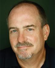
Alan G. Hinnebusch, Ph.D.
NIH Distinguished Investigator
Section on Nutrient Control of Gene Expression
NICHD/DIR
Research Topics
The goal of research in the Section on Nutrient Control of Gene Expression is to elucidate fundamental molecular mechanisms of eukaryotic gene regulation. Saccharomyces cerevisiae is employed as a model organism, allowing genetics, biochemistry, and structural biology to be combined in dissecting processes of gene transcription and mRNA translation crucial in living cells. Much work focuses on the general amino acid control, wherein amino acid biosynthetic genes in multiple pathways are induced by transcriptional activator Gcn4 in response to limitation for any amino acid. Gcn4 is one of the best-understood transcription factors regarding the structure of its activation domain, co-activator targets, and activation mechanism, its complete transcriptome, and pathways regulating its synthesis, function, and degradation. This knowledge, combined with the large battery of mutants, plasmids, and antibodies generated over the years, and new technologies based on next-generation sequencing, are being applied to dissect numerous aspects of transcriptional control of general importance in eukaryotic cells. Recent accomplishments of the group include the discovery of extensive cooperation among the chromatin remodeling complexes SWI/SNF, RSC, and Ino80C and the histone acetyltransferases (HATs) Gcn5 (in co-activator SAGA) and Eaf1 (in NuA4) in evicting and repositioning promoter nucleosomes to enable recruitment of general transcription factor TATA-binding protein (TBP) at Gcn4 target genes and other highly expressed yeast genes. They also showed that Ino80C acts in nucleosome eviction independently of its proposed function in replacing the specialized histone H2A.Z with conventional H2A in promoter nucleosomes. They discovered unexpectedly that most of the occupied Gcn4 binding sites in the yeast genome reside inside coding sequences rather than upstream of promoters; and that such non-canonical binding events frequently induce bidirectional transcription from within the coding sequences in addition to inducing the conventional full-length mRNA transcripts of the gene.
The pathway for inducing Gcn4 synthesis is another focal point of research, as this represents one branch of a dual mechanism of translational control, conserved throughout eukaryotes, that is mobilized by nutrient starvation or stress to achieve both general and gene-specific changes in protein synthesis. The intricate mechanism governing GCN4 translation entails multiple general translation initiation factors (eIFs) and a signaling pathway utilizing protein kinase Gcn2, which phosphorylates eIF2 to attenuate its function in recruiting initiator methionyl tRNA (tRNAi) to the small (40S) ribosomal subunit in response to amino acid limitation. Recently, the group identified distinct pathways for activation of kinase Gcn2 by ribosomes stalled during translation elongation that operate in the presence or absence of amino acid starvation and are, respectively, either independent or fully dependent on components of the ribosomal P-stalk.
Mutants altering GCN4 translation have been identified that affect assembly of initiation complexes, and complementary genetic selections or screens have been used to uncover mutations that alter the fidelity of selecting AUG as the initiation codon by scanning ribosomes. Analyzing the biochemical effects of such mutations in a yeast reconstituted in vitro system, in collaboration with Jon Lorsch’s group in NICHD, has illuminated in vivo mechanisms involved in recruitment of tRNAi and mRNA to ribosomes and recognition of start codons during ribosomal scanning of mRNA leader sequences. A collaboration with Venki Ramakrishnan (MRC, Cambridge) and his former colleagues Jose Llacer (Instituto de Biomedicina de Valencia) and Tanweer Hussain (Indian Institute of Science, Bangalore) has provided high-resolution structures of translation preinitiation complexes (PICs) in different states that inform the interpretations of previous findings and reveal new aspects of the initiation process that can be interrogated with genetics and biochemistry. Recent accomplishments of the group include identifying functional domains of eukaryotic initiation factors (eIFs) 1, 1A, 2, 3c, and 5, and of initiator tRNAi, 18S rRNA, and ribosomal proteins uS7/Rps5, uS3/Rps3, and uS5/Rps2 involved in accurate and efficient AUG recognition during scanning; and contributing to the elucidation of conformational transitions in the PIC through FRET analysis, chemical modification, and cryo-EM analyses of reconstituted PICs in different functional states. Other structural, biochemical and genetic evidence revealed large-scale movement of an eIF3 subdomain between solvent-exposed and subunit-interface surfaces of the 40S subunit that controls start codon recognition.
The group has identified multiple elements in yeast initiation factor eIF4G involved in activating mRNA for translation initiation, functional domains in yeast eIF4B that enhance PIC attachment to mRNA, and implicated eIF4B in eIFG∙eIF4A assembly. They defined distinct roles of DEAD-box helicases eIF4A and Ded1 (and its paralog Dbp1) in modulating translational efficiencies of mRNAs genome-wide by facilitating ribosome attachment and subsequent scanning of mRNA leaders--particularly when burdened with secondary structures. The distinct functions of eIF4A and Ded1/Dbp1 in mRNA recruitment by 48S preinitiation complexes (PICs) have been reconstituted in Lorsch's purified system, providing evidence that Ded1 interactions with eIF4A and eIF4G enhance its acceleration of 48S PIC assembly on Ded1-dependent mRNAs. They dissected the Ded1 N-terminal domain and showed that its separate interactions with eIF4A and cap-binding protein eIF4E enhance Ded1 function in vivo. They provided evidence that a Ded1-dependent mRNAs that are also highly dependent on eIF4B are translationally repressed in response to glucose starvation or heat stress.
The Section has helped to uncover functions of ABCE protein Rli1/ABCE1 in PIC assembly and, together with Rachel Green's lab, in recycling of the 60S subunit following translation termination. In collaboration with Nicholas Guydosh and Sergey Dmitriev, they further showed that yeast Tma64/Tma20/Tma22 proteins carry out the second step of ribosome recycling, dissociation of the 40S post-termination complex, and thereby block reinitiation events in 3' untranslated regions of mRNA. They further showed that impaired 40S recycling in mutants lacking the Tma factors reprograms translational efficiencies to favor efficiently translated mRNAs owing to reduced assembly of 43S PICs from recycled 40S subunits, which occurs similarly when eIF2α is phosphorylated or 40S biogenesis is impaired.
The group also established that the conserved mRNA granule protein and mRNA decapping activator Scd6 can repress translation via RNA helicase Dhh1, a known decapping activator, and accelerate decapping and mRNA degradation, when tethered to specific mRNAs in yeast cells. They further showed that Scd6, Scd6-related factor Edc3, Dhh1, and protein Pat1 collaborate to accelerate decapping of many cellular mRNAs, whereas most other transcripts decapped by the Dcp1:Dcp2 complex are targeted by nonsense-mediated decay factor Upf1. They uncovered translational repression by Dcp2 through an indirect mechanism involving suppression of mRNA abundance and a more direct mechanism involving decapping activators, and they found that decapping limits the abundance or translation of mRNAs whose products are dispensable for growth in nutrient-rich medium, including mitochondrial enzymes of respiration.
Biography
Dr. Alan G. Hinnebusch received his B.S. in Biology from the University of Dayton, Ohio, in 1975 and his Ph.D. in Biochemistry and Molecular Biology from Harvard University in 1980. He studied as a postdoctoral fellow in the laboratory of Dr. Gerald R. Fink at Cornell University and the Massachusetts Institute of Technology from 1980 to 1983. He joined the NICHD as a Senior Staff Fellow in 1983 and became Chief of the Laboratory of Eukaryotic Gene Regulation in 1995. In 2000, he was appointed as Chief of the Laboratory of Gene Regulation and Development and Head of the Section on Nutrient Control of Gene Regulation. In 2007, he was named Head of the Program in Cellular Regulation and Metabolism. Dr. Hinnebusch has served on the editorial boards of Genetics, Microbiological Reviews, Molecular Microbiology, Journal of Biological Chemistry, and Molecular & Cellular Biology, and is currently a member of the editorial boards of Genes & Development, eLife, and Genetics. He was co-organizer of the Cold Spring Harbor Laboratory Meeting on Translational Control from 2000 to 2010. He has published more than 220 original research articles in peer-reviewed journals and more than 50 review articles and book chapters pertaining to his field of research. In 1994 he was named Maryland's Outstanding Young Scientist and was elected as a Fellow of the American Academy of Microbiology. In 2009 he was elected as a Fellow of the American Association for the Advancement of Science and as a Fellow of the American Academy of Arts and Sciences, and in 2015 he was elected to the National Academy of Sciences.
Selected Publications
- Rawal Y, Chereji RV, Valabhoju V, Qiu H, Ocampo J, Clark DJ, Hinnebusch AG. Gcn4 Binding in Coding Regions Can Activate Internal and Canonical 5' Promoters in Yeast. Mol Cell. 2018;70(2):297-311.e4.
- Qiu H, Biernat E, Govind CK, Rawal Y, Chereji RV, Clark DJ, Hinnebusch AG. Chromatin remodeler Ino80C acts independently of H2A.Z to evict promoter nucleosomes and stimulate transcription of highly expressed genes in yeast. Nucleic Acids Res. 2020;48(15):8408-8430.
- Vijjamarri AK, Niu X, Vandermeulen MD, Onu C, Zhang F, Qiu H, Gupta N, Gaikwad S, Greenberg ML, Cullen PJ, Lin Z, Hinnebusch AG. Decapping factor Dcp2 controls mRNA abundance and translation to adjust metabolism and filamentation to nutrient availability. Elife. 2023;12.
- Gulay S, Gupta N, Lorsch JR, Hinnebusch AG. Distinct interactions of eIF4A and eIF4E with RNA helicase Ded1 stimulate translation in vivo. Elife. 2020;9.
- Gupta R, Hinnebusch AG. Differential requirements for P stalk components in activating yeast protein kinase Gcn2 by stalled ribosomes during stress. Proc Natl Acad Sci U S A. 2023;120(16):e2300521120.
Related Scientific Focus Areas
Molecular Biology and Biochemistry
View additional Principal Investigators in Molecular Biology and Biochemistry
Microbiology and Infectious Diseases
View additional Principal Investigators in Microbiology and Infectious Diseases
This page was last updated on Monday, October 30, 2023