Eric Betzig’s Nobel Prize, a homegrown success
A letter from Michael M. Gottesman, Deputy Director for Intramural Research
Dear colleagues,
The NIH intramural program has placed its mark on another Nobel Prize. You likely heard last week that Eric Betzig of HHMI’s Janelia Farm Research Campus will share the 2014 Nobel Prize in Chemistry “for the development of super-resolved fluorescence microscopy.” Eric's key experiment came to life right here at the NIH, in the lab of Jennifer Lippincott-Schwartz.
In fact, Eric’s story is quite remarkable and highlights the key strengths of our intramural program: freedom to pursue high-risk research, opportunities to collaborate, and availability of funds to kick-start such a project.
Eric was “homeless” from a scientist’s viewpoint. He was unemployed and working out of a cottage in rural Michigan with no way of turning his theory into reality. He had a brilliant idea to isolate individual fluorescent molecules by a unique optical feature to overcome the diffraction limit of light microscopes, which is about 0.2 microns. He thought that if green fluorescent proteins (GFPs) could be switched on and off a few molecules at a time, it might be possible using Gaussian fitting to synthesize a series of images based on point localization that, when stacked, provide extraordinary resolution.

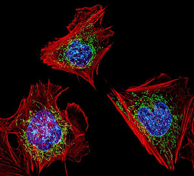
Credit: Dylan Burnette and Jennifer Lippincott-Schwartz, Eunice Kennedy Shriver National Institute of Child Health and Human Development, National Institutes of Health
The cells shown here are fibroblasts, one of the most common cells in mammalian connective tissue. These particular cells were taken from a mouse. Scientists used them to test the power of a new microscopy technique that offers vivid views of the inside of a cell. The DNA within the nucleus (blue), mitochondria (green) and cellular skeleton (red) is clearly visible.
Eric chanced to meet Jennifer, who heads the NICHD’s Section on Organelle Biology. She and George Patterson, then a postdoc in Jennifer’s lab and now a PI in NIBIB, had developed a photoactivable version of GFP with these capabilities, which they were already applying to the study of organelles. Jennifer latched on to Eric’s idea immediately; she was among the first to understand its significance and saw that her laboratory had just the tool that Eric needed.
So, in mid-2005, Jennifer offered to host Eric and his friend and colleague, Harald Hess, to collaborate on building a super-resolution microscope based on the use of photoactivatable GFP. The two had constructed key elements of this microscope in Harald's living room out of their personal funds.
Jennifer located a small space in her lab in Building 32. She and Juan Bonifacino, also in NICHD, then secured some centralized IATAP funds for microscope parts to supplement the resources that Eric and Harald brought to the lab. Owen Rennert, then the NICHD scientific director, provided matching funds. By October 2005, Eric and Harald became affiliated with HHMI, which also contributed funds to the project.
Eric and Harald quickly got to work with their new NICHD colleagues in their adopted NIH home. The end result was a fully operational microscope married to GFP technology capable of producing super-resolution images of intact cells for the first time. Called photoactivated localization microscopy (PALM), the new technique provided 10 times the resolution of conventional light microscopy.
Another postdoc in Jennifer’s lab, Rachid Sougrat, now at King Abdullah University of Science and Technology in Saudi Arabia, correlated the PALM images of cell organelles to electron micrographs to validate the new technique, yet another important contribution.
Upon hearing of Eric’s Nobel Prize, Jennifer told me: “We didn’t imagine at the time how quickly the point localization imaging would become such an amazing enabling technology; but it caught on like wildfire, expanding throughout many fields of biology.”
That it did! PALM and all its manifestations are at the heart of extraordinary discoveries. We think this is a quintessential intramural story. We see the elements of high-risk/high-reward research and the importance of collaboration and the freedom to pursue ideas, as well as NIH scientists with the vision to encourage and support this research.
Read the landmark 2006 Science article by Eric, Harald, and the NICHD team, “Imaging Intracellular Fluorescent Proteins at Nanometer Resolution,” at http://www.sciencemag.org/content/313/5793/1642.long.
The story of the origins of Eric Betzig’s Nobel Prize in Jennifer Lippincott-Schwartz’s lab is one that needs to be told. I feel proud to work for an organization that can attract such talent and enable such remarkable science to happen.
Kudos to Eric and to Jennifer and her crew.
Michael M. Gottesman
Deputy Director for Intramural Research
Read more about their exciting story at the following links:
https://irp.nih.gov/our-research/research-in-action/seeing-is-believing
https://irp.nih.gov/catalyst/v21i5/how-cells-crawl
This page was last updated on Thursday, January 20, 2022